
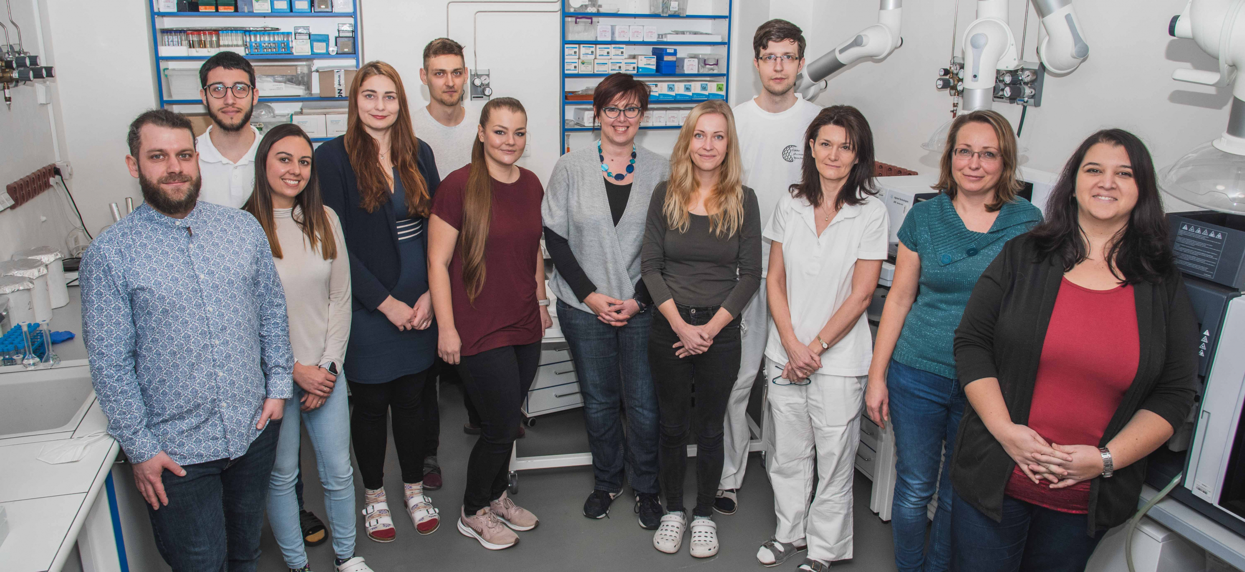
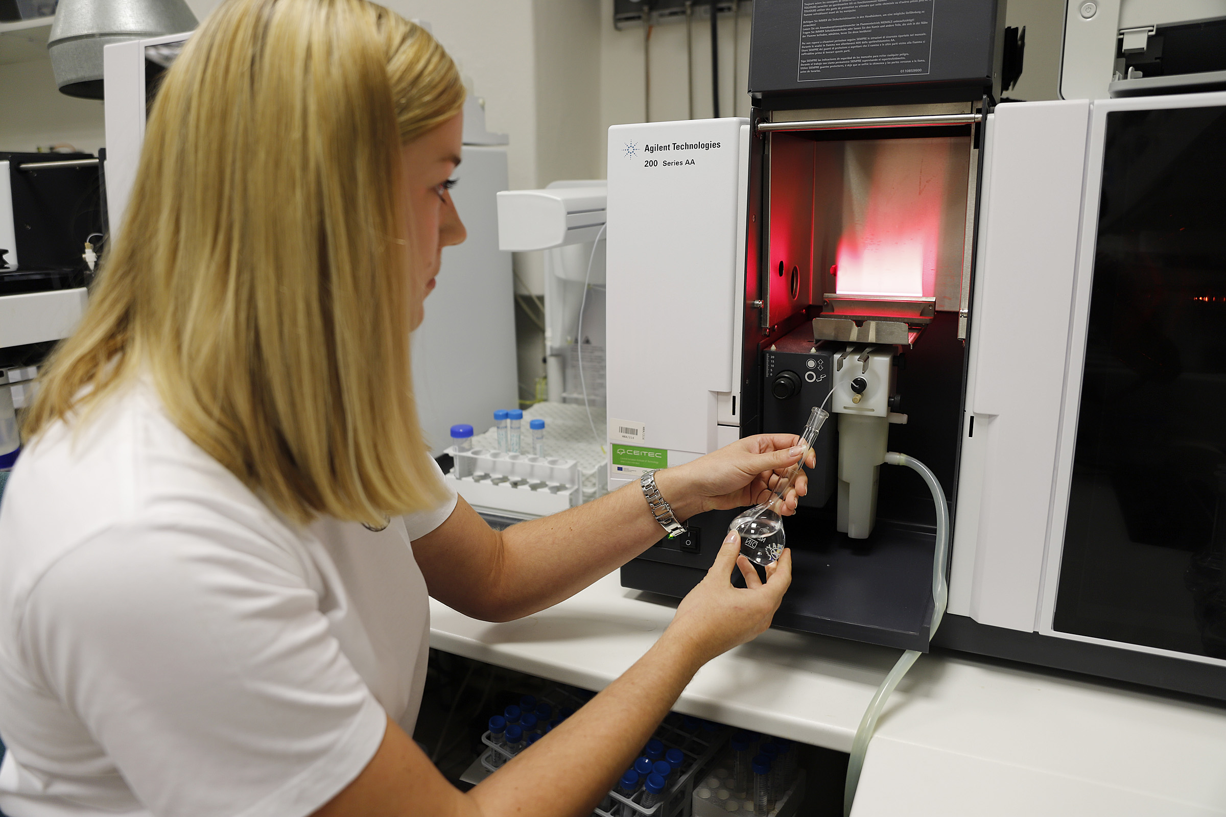
Výzkumná skupina instrumentální analýzy a bioanalýzy
Multidisciplinární výzkumná skupina se zaměřuje na široké spektrum vědeckých témat pokrývajících oblasti analytické chemie, elementární analýzy a bioanalýzy. Nově vyvinuté nebo optimalizované metody jsou uplatněny nejen v oblasti konvenčních aplikací (environmentální analýza, analýza potravin, medicinská diagnostika, aj.) ale také pro nalezení důležitých biologických souvislostí nebo monitorování vlivu externích faktorů na klíčové biologické procesy.
Využitím nejen špičkové analytické instrumentace (spektrometrické nebo separační techniky) ale také špičkových postupů moderní molekulární biologie (editace genomu) je dosaženo komplexního přístupu.
Jednou z hlavních výhod multidisciplinárního a multiinstrumentálního přístupu je možnost získat informace nejen o obsahu kovů (AAS, (LA)-ICP-MS) ale také klíčových biomolekul (DART/DESI-MS, GC-MS).
Výzkumná skupina má nejen širokou síť tuzemských i mezinárodních spolupracovníků, ale provádí i zakázkovou činnost a to především v oblasti stanovení kovů a jejich speciace.


doc. Mgr. Markéta Vaculovičová, Ph.D.
Vedoucí výzkumné skupiny instrumentální analýzy a bioanalýzy
Akademická pracovnice – docentka
Telefon: 420545 13 32 90
Adresa pracoviště: ÚCB AF, Zemědělská 1, 61300 Brno – Budova D
Označení kanceláře: BA02N3010
E-mail: Marketa.Ryvolova@seznam.cz
Researcher ID: E-5583-2016
ORCID: 0000-0002-6771-1304
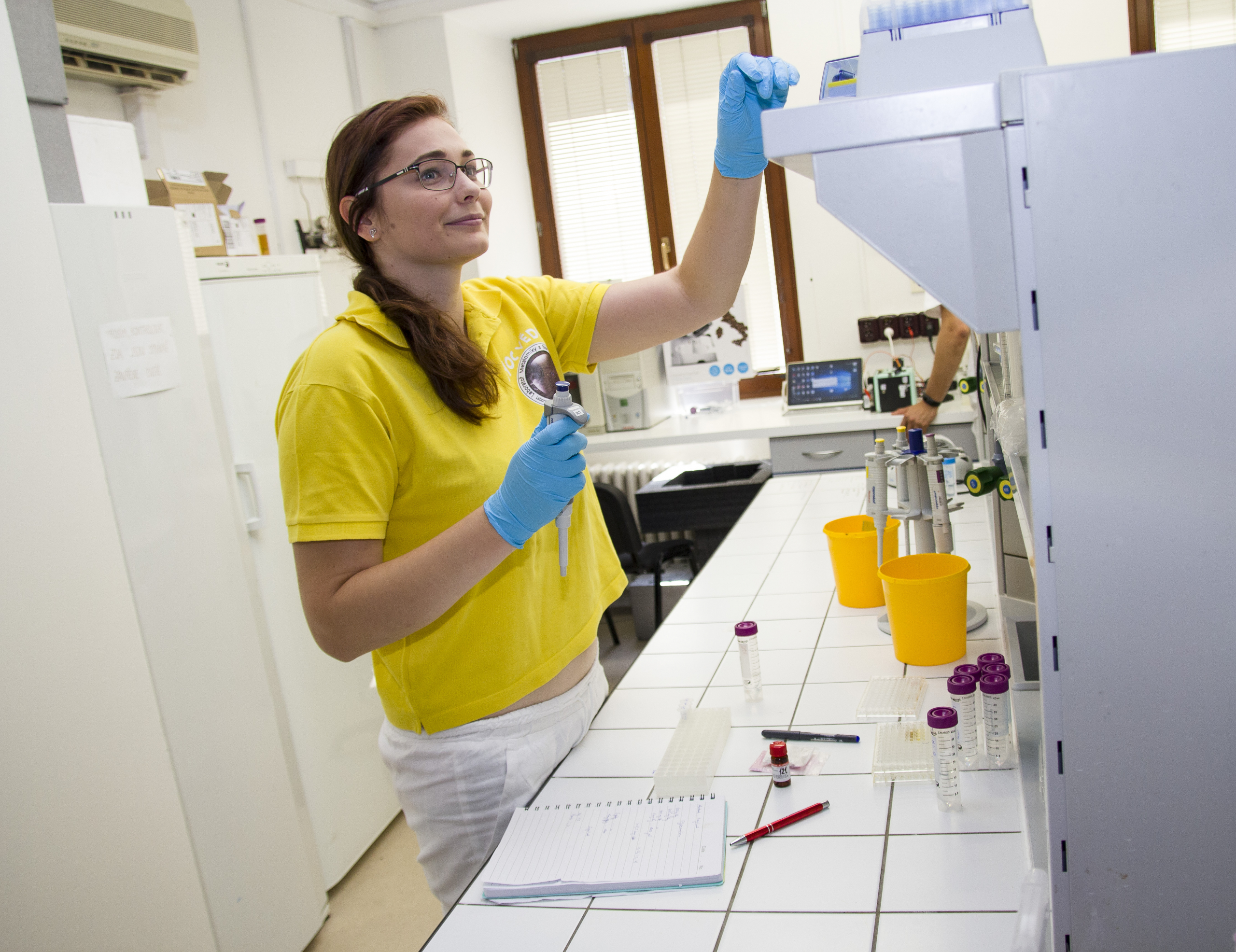
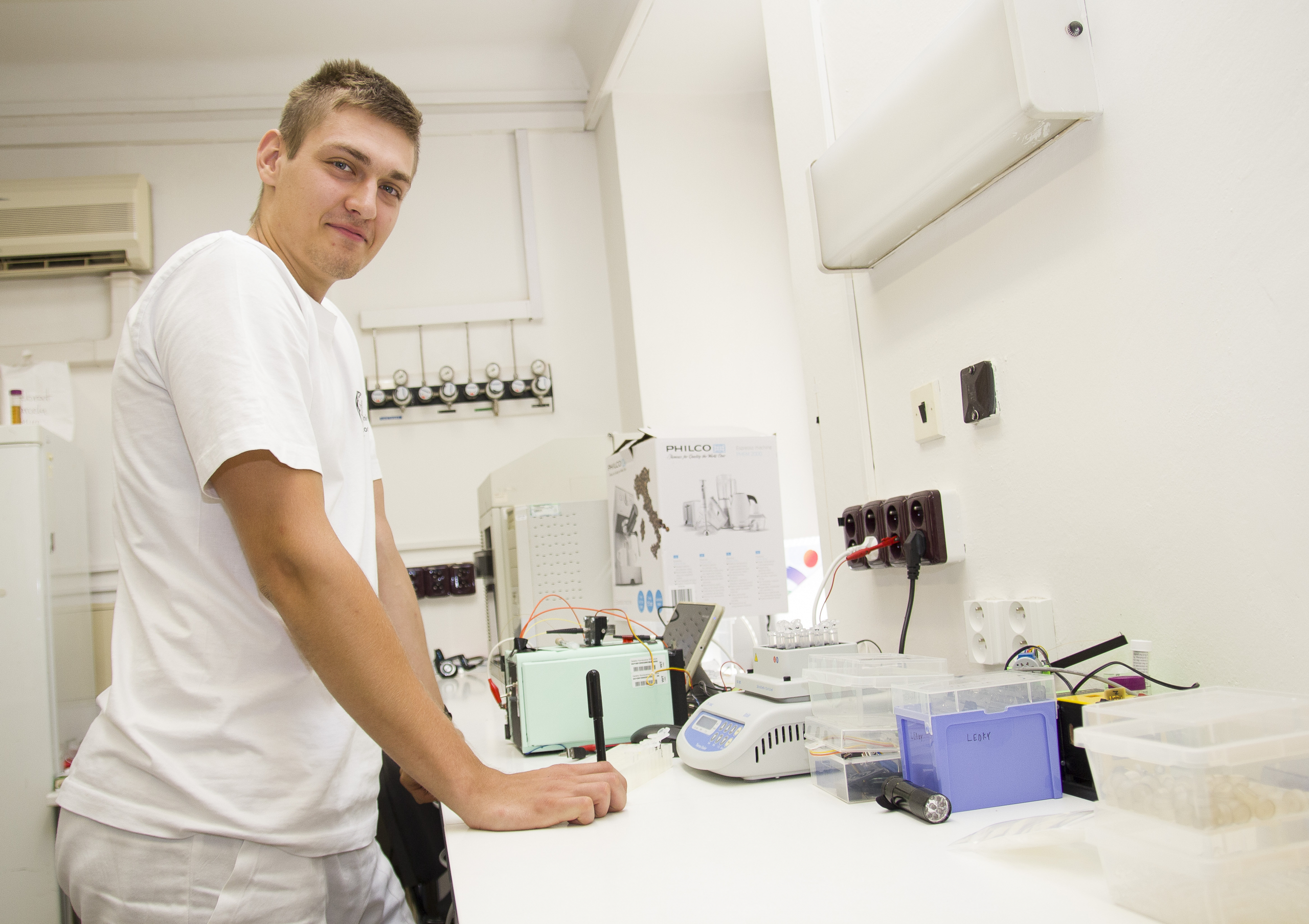
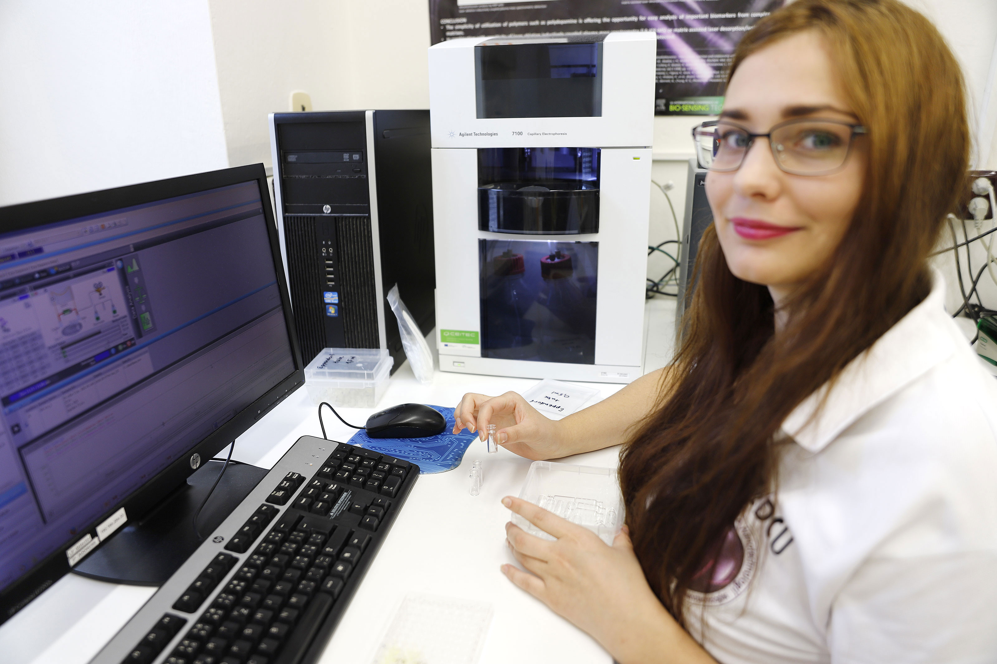
Členové týmu
- doc. Mgr. Markéta Vaculovičová, Ph.D.
- Ing. Lukáš Nejdl, Ph.D.
- Ing., Andrea Ridošková, Ph.D.
- doc. Mgr. Pavlína Pelcová, Ph.D.
- prof. RNDr. Miroslav Macka, Ph.D.
- Mgr. Dagmar Štěrbová, Ph.D.
- Ing. Jaroslava Bezděková, Ph.D.
- Ing. Kristýna Pavelicová, Ph.D.
- Ing. Vendula Smolíková, Ph.D.
- Ing. Kristýna Veverková, Ph.D.
- Mgr. Tomáš Rýpar
- Mgr. Marcela Vlčnovská
- Ing. et Ing. Milada Vodová
- Peter Csányi
Média
- Česká televize – Takzvaný „UV otisk prstu“ přináší nečekané možnosti nejen kriminalistům
- Lukáš Nejdl: Věda je velké dobrodružství, které mění sny v realitu
- O vědě a vědcích: Analýza paprskem UV-C
- Pančování vína či původ drog rozeznají brněnští vědci pomocí ÚV záření
Vypsaná témata disertačních prací
- Volné téma-kovy v životním prostředí
- Biologická dostupnost rtuti pro užitkové plodiny
- Parefluidní analytická zařízení pro využití v diagnostice
- Optické detekční techniky pro využití v bioanalýze
- Vývoj nových přístupů pro klinickou diagnostiku
- Vývoj biosenzorů pro rychlou a jednoduchou diagnostiku
- Volné téma
Projekty
- AZV ČR: Role signalizace monocytárních buněk, jejich metabolických změn a transkripčních faktorů u pacientů se závažnou sepsí.. 2018-2021.
- FAO/IAEA: Kutikulární uhlovodíky: Nový chemotaxonomický a chemoekologický
nástroj pro kontrolu zemědělských škůdců vrtulí z rodů Zeugodacus a Bactrocera. 2019-2023. - GAČR: Paperfluidická přenosná zařizení pro rychlou a nízkonákladovou analýzu bez instrumentální detekce. 2019-2021.
Publikace
2019
Rahbar, Mohammad; Wheeler, Aaron R; Paull, Brett; Macka, Mirek
Ion-Exchange Based Immobilization of Chromogenic Reagents on Microfluidic Paper Analytical Devices Journal Article
In: Anal Chem, vol. 91, no. 14, pp. 8756–8761, 2019, ISSN: 1520-6882.
@article{pmid31251584,

title = {Ion-Exchange Based Immobilization of Chromogenic Reagents on Microfluidic Paper Analytical Devices},

author = {Mohammad Rahbar and Aaron R Wheeler and Brett Paull and Mirek Macka},

doi = {10.1021/acs.analchem.9b01288},

issn = {1520-6882},

year  = {2019},

date = {2019-01-01},

journal = {Anal Chem},

volume = {91},

number = {14},

pages = {8756--8761},

abstract = {Distance-based detection methods, as used in development of microfluidic paper analytical devices (μPADs), rely on the dynamic formation of a colored band along the length of the paper microfluidic channels. The color change is driven by the reaction of chromogenic reagents (typically water-insoluble) that are bound to the paper, thus not subject to being washed away by the sample flow along the detection channel. Here, we introduce the use of an anion-exchange filter paper (as a replacement for standard, unmodified filter paper) for distance-based detection in μPADs, in order to immobilize the water-soluble anionic reagents upon the paper detection channels based on ion-exchange interactions of the oppositely charged paper (protonated tertiary amine groups) and the anionic groups of the reagents. The ion-exchange (IE) paper was initially characterized and its properties were compared with standard cellulose paper. The IE paper was shown to be capable of strong retention of anionic reagents exhibiting acidic functional groups (carboxylic, sulfonic), which become deprotonated and negatively charged when in contact with the IE paper. The effect of the ionic strength (10-250 mM Cl) and pH (1-13) on the immobilization of the investigated reagents were also determined. The IE-μPADs were then modified with anionic chromogenic reagents and applied to distance-based determination of total calcium (LOD = 0.03 mM) and total acidity (LOD = 2.5 mM) content in serum and wine samples, respectively. The detailed mechanisms of the developed assays on the IE paper are also discussed. We propose that IE-μPADs represent a useful new addition to the distance-based detection toolbox and considerably enhance the applicability of such a detection method.},

keywords = {},

pubstate = {published},

tppubtype = {article}

}
Rahbar, Mohammad; Nesterenko, Pavel N; Paull, Brett; Macka, Mirek
High-throughput deposition of chemical reagents via pen-plotting technique for microfluidic paper-based analytical devices Journal Article
In: Anal Chim Acta, vol. 1047, pp. 115–123, 2019, ISSN: 1873-4324.
@article{pmid30567641,

title = {High-throughput deposition of chemical reagents via pen-plotting technique for microfluidic paper-based analytical devices},

author = {Mohammad Rahbar and Pavel N Nesterenko and Brett Paull and Mirek Macka},

doi = {10.1016/j.aca.2018.09.006},

issn = {1873-4324},

year  = {2019},

date = {2019-01-01},

journal = {Anal Chim Acta},

volume = {1047},

pages = {115--123},

abstract = {The deposition of chemical reagent inks on paper is a crucial step in the development and fabrication of microfluidic paper-based analytical devices (μPADs). A pen-plotting approach, delivering chemical ink deposition using technical pens filled with reagents and inserted into a desktop electronic plotter, is shown herein to be a versatile, low-cost, simple, rapid, reproducible, and high-throughput solution. The volume of the deposited ink was quantified gravimetrically, confirming that nanoliter volumes of reagents can be deposited reproducibly (e.g. 7.55 ± 0.37 nL/mm for a plotting speed of 10 cm/s) in detection zones of μPADs, typically spatially defined using wax printing. This approach was further investigated with regard to deposition of reagents in different geometrical forms (circular and linear), so demonstrating its applicability for preparation of μPADs with flexible design and application. By adjusting the plotting speed for linear deposition, lines with a relatively large range of widths (≈628-1192 μm) were created. Circular deposition was also demonstrated via delivery of reagents within wax printed circular fluidic barriers of a range of diameters (inner diameter = 1.5-7 mm). These capabilities were practically demonstrated via the fabrication of μPADs, based upon differing detection principles for determination of aluminum in natural waters using Morin as the fluorescent reagent. Traditional μPADs based on digital image colorimetry (DIC) were produced using circular deposition, whilst distance-based μPADs exploited linear deposition. Both types of μPADs developed using this method showed excellent precision for determination of trace concentrations of aluminium (average RSDs = 3.38% and 6.45%, and LODs = 0.5 ng (0.25 ppm) and 2 ng (0.5 ppm), for traditional and distance-based detection, respectively).},

keywords = {},

pubstate = {published},

tppubtype = {article}

}
Vaneckova, Tereza; Bezdekova, Jaroslava; Tvrdonova, Michaela; Vlcnovska, Marcela; Novotna, Veronika; Neuman, Jan; Stossova, Aneta; Kanicky, Viktor; Adam, Vojtech; Vaculovicova, Marketa; Vaculovic, Tomas
In: Sci Rep, vol. 9, no. 1, pp. 11840, 2019, ISSN: 2045-2322.
@article{pmid31413275,

title = {CdS quantum dots-based immunoassay combined with particle imprinted polymer technology and laser ablation ICP-MS as a versatile tool for protein detection},

author = {Tereza Vaneckova and Jaroslava Bezdekova and Michaela Tvrdonova and Marcela Vlcnovska and Veronika Novotna and Jan Neuman and Aneta Stossova and Viktor Kanicky and Vojtech Adam and Marketa Vaculovicova and Tomas Vaculovic},

doi = {10.1038/s41598-019-48290-2},

issn = {2045-2322},

year  = {2019},

date = {2019-01-01},

journal = {Sci Rep},

volume = {9},

number = {1},

pages = {11840},

abstract = {For the first time, the combination of molecularly imprinted polymer (MIP) technology with laser ablation inductively coupled plasma mass spectrometry (LA-ICP-MS) is presented with focus on an optimization of the LA-ICP-MS parameters such as laser beam diameter, laser beam fluence, and scan speed using CdS quantum dots (QDs) as a template and dopamine as a functional monomer. A non-covalent imprinting approach was employed in this study due to the simplicity of preparation. Simple oxidative polymerization of the dopamine that creates the self-assembly monolayer seems to be an ideal choice. The QDs prepared by UV light irradiation synthesis were stabilized by using mercaptosuccinic acid. Formation of a complex of QD-antibody and QD-antibody-antigen was verified by using capillary electrophoresis with laser-induced fluorescence detection. QDs and antibody were connected together via an affinity peptide linker. LA-ICP-MS was employed as a proof-of-concept for detection method of two types of immunoassay: 1) antigen extracted from the sample by MIP and subsequently overlaid/immunoreacted by QD-labelled antibodies, 2) complex of antigen, antibody, and QD formed in the sample and subsequently extracted by MIP. The first approach provided higher sensitivity (MIP/NIP), however, the second demonstrated higher selectivity. A mixture of proteins with size in range 10-250 kDa was used as a model sample to demonstrate the capability of both approaches for detection of IgG in a complex sample.},

keywords = {},

pubstate = {published},

tppubtype = {article}

}
Huang, Ling; Kakadiaris, Eugenia; Vaneckova, Tereza; Huang, Kai; Vaculovicova, Marketa; Han, Gang
Designing next generation of photon upconversion: Recent advances in organic triplet-triplet annihilation upconversion nanoparticles Journal Article
In: Biomaterials, vol. 201, pp. 77–86, 2019, ISSN: 1878-5905.
@article{pmid30802685,

title = {Designing next generation of photon upconversion: Recent advances in organic triplet-triplet annihilation upconversion nanoparticles},

author = {Ling Huang and Eugenia Kakadiaris and Tereza Vaneckova and Kai Huang and Marketa Vaculovicova and Gang Han},

doi = {10.1016/j.biomaterials.2019.02.008},

issn = {1878-5905},

year  = {2019},

date = {2019-01-01},

journal = {Biomaterials},

volume = {201},

pages = {77--86},

abstract = {Organic triplet-triplet annihilation upconversion (TTA-UC) nanoparticles have emerged as exciting therapeutic agents and imaging probes in recent years due to their unique chemical and optical properties such as outstanding biocompatibility and low power excitation density. In this review, we focus on the latest breakthroughs in such new version of upconversion nanoparticle, including their design, preparation, and applications. First, we will discuss the key principles and design concept of these organic-based photon upconversion in regard to the methods of selection of the related triplet TTA dye pairs (photosensitizer and emitter). Then, we will discuss the recent approaches s to construct TTA-UCNPs including silica TTA-UCNPs, lipid-coated TTA-UCNPs, polymer encapsulated TTA-UCNPs, nano-droplet TTA-UCNPs and metal-organic frameworks (MOFs) constructed TTA-UCNPs. In addition, the applications of TTA-UCNPs will be discussed. Finally, we will discuss the challenges posed by current TTA-UCNP development.},

keywords = {},

pubstate = {published},

tppubtype = {article}

}
Jelinkova, Pavlina; Mazumdar, Aninda; Sur, Vishma Pratap; Kociova, Silvia; Dolezelikova, Kristyna; Jimenez, Ana Maria Jimenez; Koudelkova, Zuzana; Mishra, Pawan Kumar; Smerkova, Kristyna; Heger, Zbynek; Vaculovicova, Marketa; Moulick, Amitava; Adam, Vojtech
Nanoparticle-drug conjugates treating bacterial infections Journal Article
In: J Control Release, vol. 307, pp. 166–185, 2019, ISSN: 1873-4995.
@article{pmid31226356,

title = {Nanoparticle-drug conjugates treating bacterial infections},

author = {Pavlina Jelinkova and Aninda Mazumdar and Vishma Pratap Sur and Silvia Kociova and Kristyna Dolezelikova and Ana Maria Jimenez Jimenez and Zuzana Koudelkova and Pawan Kumar Mishra and Kristyna Smerkova and Zbynek Heger and Marketa Vaculovicova and Amitava Moulick and Vojtech Adam},

doi = {10.1016/j.jconrel.2019.06.013},

issn = {1873-4995},

year  = {2019},

date = {2019-01-01},

journal = {J Control Release},

volume = {307},

pages = {166--185},

abstract = {The ever increasing scenario of bacterial resistance against commonly available antibiotics is becoming a global threat of major concern, which necessitates the development of new strategies to overcome this hurdle. Conjugation of nanoparticles (NPs) with antimicrobial moieties, such as antibiotics, peptides or different biomolecules, has been one of the successful techniques in targeting antibiotic resistance. This review mainly focusses on the possible nanoparticle-drug conjugates with their activity against pathogenic bacterial infections. Nanoparticles play an array of roles, e.g. as a carrier, synergistically acting agent and as theranostic agent, henceforth facilitates the efficacy of therapy. Moreover, this review elaborates the studies with reported nanoparticles-drug conjugates that include their possible synthesis methodologies and applications. In most of the cases, the nanoparticles were found to increase the permeability of bacterial cell membrane, which enables higher uptake of antibiotics inside the bacterial cells which in return showed better effects. Even the conjugates were found to efficiently kill the antibiotic-resistant strains. Since several limitations are exerted by the biological systems, there is an urge for the advancement of nanoparticle-drug conjugates for better proficiency.},

keywords = {},

pubstate = {published},

tppubtype = {article}

}
Tvrdonova, Michaela; Vlcnovska, Marcela; Vanickova, Lucie Pompeiano; Kanicky, Viktor; Adam, Vojtech; Ascher, Lena; Jakubowski, Norbert; Vaculovicova, Marketa; Vaculovic, Tomas
Gold nanoparticles as labels for immunochemical analysis using laser ablation inductively coupled plasma mass spectrometry Journal Article
In: Anal Bioanal Chem, vol. 411, no. 3, pp. 559–564, 2019, ISSN: 1618-2650.
@article{pmid30109381,

title = {Gold nanoparticles as labels for immunochemical analysis using laser ablation inductively coupled plasma mass spectrometry},

author = {Michaela Tvrdonova and Marcela Vlcnovska and Lucie Pompeiano Vanickova and Viktor Kanicky and Vojtech Adam and Lena Ascher and Norbert Jakubowski and Marketa Vaculovicova and Tomas Vaculovic},

doi = {10.1007/s00216-018-1300-7},

issn = {1618-2650},

year  = {2019},

date = {2019-01-01},

journal = {Anal Bioanal Chem},

volume = {411},

number = {3},

pages = {559--564},

abstract = {In this paper, we describe the labelling of antibodies by gold nanoparticles (AuNPs) with diameters of 10 and 60 nm with detection by laser ablation inductively coupled plasma mass spectrometry (LA-ICP-MS). Additionally, the AuNPs labelling strategy is compared with commercially available labelling reagents based on MeCAT (metal coded affinity tagging). Proof of principle experiments based on dot blot experiments were performed. The two labelling methods investigated were compared by sensitivity and limit of detection (LOD). The absolute LODs achieved were in the range of tens of picograms for AuNP labelling compared to a few hundred picograms by the MeCAT labelling.},

keywords = {},

pubstate = {published},

tppubtype = {article}

}
Strmiska, Vladislav; Michalek, Petr; Lackova, Zuzana; Guran, Roman; Krizkova, Sona; Vanickova, Lucie; Zitka, Ondrej; Stiborova, Marie; Eckschlager, Tomas; Klejdus, Borivoj; Pacik, Dalibor; Tvrdikova, Eliska; Keil, Claudia; Haase, Hajo; Adam, Vojtech; Heger, Zbynek
Sarcosine is a prostate epigenetic modifier that elicits aberrant methylation patterns through the SAMe-Dnmts axis Journal Article
In: Mol Oncol, vol. 13, no. 5, pp. 1002–1017, 2019, ISSN: 1878-0261.
@article{pmid30628163,

title = {Sarcosine is a prostate epigenetic modifier that elicits aberrant methylation patterns through the SAMe-Dnmts axis},

author = {Vladislav Strmiska and Petr Michalek and Zuzana Lackova and Roman Guran and Sona Krizkova and Lucie Vanickova and Ondrej Zitka and Marie Stiborova and Tomas Eckschlager and Borivoj Klejdus and Dalibor Pacik and Eliska Tvrdikova and Claudia Keil and Hajo Haase and Vojtech Adam and Zbynek Heger},

doi = {10.1002/1878-0261.12439},

issn = {1878-0261},

year  = {2019},

date = {2019-01-01},

journal = {Mol Oncol},

volume = {13},

number = {5},

pages = {1002--1017},

abstract = {DNA hypermethylation is one of the most common epigenetic modifications in prostate cancer (PCa). Several studies have delineated sarcosine as a PCa oncometabolite that increases the migration of malignant prostate cells while decreasing their doubling time. Here, we show that incubation of prostate cells with sarcosine elicited the upregulation of sarcosine N-demethylation enzymes, sarcosine dehydrogenase and pipecolic acid oxidase. This process was accompanied by a considerable increase in the production of the major methyl-donor S-adenosylmethionine (SAMe), together with an elevation of cellular methylation potential. Global DNA methylation analyses revealed increases in methylated CpG islands in distinct prostate cell lines incubated with sarcosine, but not in cells of nonprostate origin. This phenomenon was further associated with marked upregulation of DNA methyltransferases (Dnmts). Epigenetic changes were recapitulated through blunting of Dnmts using the hypomethylating agent 5-azacytidine, which was able to inhibit sarcosine-induced migration of prostate cells. Moreover, spatial mapping revealed concomitant increases in sarcosine, SAMe and Dnmt1 in histologically confirmed malignant prostate tissue, but not in adjacent or nonmalignant tissue, which is in line with the obtained in vitro data. In summary, we show here for the first time that sarcosine acts as an epigenetic modifier of prostate cells and that this may contribute to its oncometabolic role.},

keywords = {},

pubstate = {published},

tppubtype = {article}

}
Ridošková, Andrea; Pelfrêne, Aurélie; Douay, Francis; Pelcová, Pavlína; Smolíková, Vendula; Adam, Vojtěch
In: Environ Toxicol Chem, vol. 38, no. 2, pp. 321–328, 2019, ISSN: 1552-8618.
@article{pmid30620786,

title = {Bioavailability of mercury in contaminated soils assessed by the diffusive gradient in thin film technique in relation to uptake by Miscanthus × giganteus},

author = {Andrea Ridošková and Aurélie Pelfrêne and Francis Douay and Pavlína Pelcová and Vendula Smolíková and Vojtěch Adam},

doi = {10.1002/etc.4318},

issn = {1552-8618},

year  = {2019},

date = {2019-01-01},

journal = {Environ Toxicol Chem},

volume = {38},

number = {2},

pages = {321--328},

abstract = {We assessed the relationship between the diffusive gradient in thin film (DGT) technique using the new ion-exchange resin Ambersep GT74 and the uptake of mercury (Hg) by a model plant cultivated on metal-contaminated agricultural soils under greenhouse conditions. Based on the total Hg content, 0.37 to 1.17% of the Hg passed to the soil porewater from the solid phase, and 2.18 to 9.18% of the Hg is DGT-available. These results were confirmed by calculating the R value (the ratio of the concentrations of bioavailable Hg measured by DGT and soil solution), which illustrated the strong bonding of Hg to the solid phase of soil and its extremely low mobility. Only inorganic Hg species were found in the metal-contaminated agricultural soils, as determined by a high-performance liquid chromatography-cold vapor atomic fluorescence spectrometry speciation analysis. The Hg was distributed in Miscanthus × giganteus organs in the following order for all sampling sites: roots (55-82%) >> leaves (8-27%) > stems (7-16%) > rhizomes (4-7%). Environ Toxicol Chem 2019;38:321-328. © 2018 SETAC.},

keywords = {},

pubstate = {published},

tppubtype = {article}

}
Ridošková, Andrea; Pelfrêne, Aurélie; Douay, Francis; Pelcová, Pavlína; Smolíková, Vendula; Adam, Vojtěch
In: Environ Toxicol Chem, vol. 38, no. 2, pp. 321–328, 2019, ISSN: 1552-8618.
@article{pmid30620786b,

title = {Bioavailability of mercury in contaminated soils assessed by the diffusive gradient in thin film technique in relation to uptake by Miscanthus × giganteus},

author = {Andrea Ridošková and Aurélie Pelfrêne and Francis Douay and Pavlína Pelcová and Vendula Smolíková and Vojtěch Adam},

doi = {10.1002/etc.4318},

issn = {1552-8618},

year  = {2019},

date = {2019-01-01},

journal = {Environ Toxicol Chem},

volume = {38},

number = {2},

pages = {321--328},

abstract = {We assessed the relationship between the diffusive gradient in thin film (DGT) technique using the new ion-exchange resin Ambersep GT74 and the uptake of mercury (Hg) by a model plant cultivated on metal-contaminated agricultural soils under greenhouse conditions. Based on the total Hg content, 0.37 to 1.17% of the Hg passed to the soil porewater from the solid phase, and 2.18 to 9.18% of the Hg is DGT-available. These results were confirmed by calculating the R value (the ratio of the concentrations of bioavailable Hg measured by DGT and soil solution), which illustrated the strong bonding of Hg to the solid phase of soil and its extremely low mobility. Only inorganic Hg species were found in the metal-contaminated agricultural soils, as determined by a high-performance liquid chromatography-cold vapor atomic fluorescence spectrometry speciation analysis. The Hg was distributed in Miscanthus × giganteus organs in the following order for all sampling sites: roots (55-82%) >> leaves (8-27%) > stems (7-16%) > rhizomes (4-7%). Environ Toxicol Chem 2019;38:321-328. © 2018 SETAC.},

keywords = {},

pubstate = {published},

tppubtype = {article}

}
2018
Li, Yan; Nesterenko, Pavel N; Stanley, Roger; Paull, Brett; Macka, Mirek
High sensitivity deep-UV LED-based z-cell photometric detector for capillary liquid chromatography Journal Article
In: Anal Chim Acta, vol. 1032, pp. 197–202, 2018, ISSN: 1873-4324.
@article{pmid30143218,

title = {High sensitivity deep-UV LED-based z-cell photometric detector for capillary liquid chromatography},

author = {Yan Li and Pavel N Nesterenko and Roger Stanley and Brett Paull and Mirek Macka},

doi = {10.1016/j.aca.2018.06.005},

issn = {1873-4324},

year  = {2018},

date = {2018-11-01},

journal = {Anal Chim Acta},

volume = {1032},

pages = {197--202},

abstract = {A new high sensitivity deep-UV LED photometric detector with a z-type flow cell (45 nL or 180 nL) for miniaturised and portable capillary liquid chromatography (LC) was designed and fabricated to overcome sensitivity limitations due to short pathlength in on-capillary detectors. The new detector has a 10 mm geometric pathlength and uses high intensity light-emitting diodes (LED) as light sources in the deep-UV range (254 nm and 280 nm). No optical reference was necessary due to the low drift in the signal. Stray light was minimized by the use of an adjustable slit with a 0.5 mm pinhole. The direct relationship between absorbance and concentration was obtained using dichromate to evaluate the sensitivity and the linearity range of the detector. Performance of the miniaturised version was compared with that obtained from a commercial benchtop detector for capillary LC under the same conditions using the same optical z-cell. The miniaturised version exhibited a superior performance across all parameters, including 3 times higher effective pathlength, 4 times higher upper limit of detector linearity, and 2-5 times lower stray light levels. An application of the new detector was shown with the detection of l-dopa, l-tyrosine, norfenefrine, phenylephrine and tyramine, separated using capillary LC. The baseline noise level recorded was as low as 3.9 μAU. Further, the detector was applied in a miniaturised capillary LC for the indirect detection of common inorganic anions. In comparison to an on-capillary LED detector applied under similar chromatographic conditions, there was a 50 times higher signal to noise (S/N) ratio.},

keywords = {},

pubstate = {published},

tppubtype = {article}

}
Pelcová, Pavlína; Vičarová, Petra; Dočekalová, Hana; Poštulková, Eva; Kopp, Radovan; Mareš, Jan; Smolíková, Vendula
In: Chemosphere, vol. 211, pp. 1109–1112, 2018, ISSN: 1879-1298.
@article{pmid30223326,

title = {The prediction of mercury bioavailability for common carp (Cyprinus carpio L.) using the DGT technique in the presence of chloride ions and humic acid},

author = {Pavlína Pelcová and Petra Vičarová and Hana Dočekalová and Eva Poštulková and Radovan Kopp and Jan Mareš and Vendula Smolíková},

doi = {10.1016/j.chemosphere.2018.07.202},

issn = {1879-1298},

year  = {2018},

date = {2018-11-01},

journal = {Chemosphere},

volume = {211},

pages = {1109--1112},

abstract = {The ability of the DGT technique to predict Hg bioavailability for input tissues (skin, gills, and scales) of common carp in the presence of chloride ions and humic acid (HA) was evaluated. The mercury accumulation by the DGT units and input tissues of carp decreased with an increasing concentration of chloride ions (29-180 mg L) and HA (0-5 mg L). In the presence of chloride ions and HA, statistically significant correlations (Pearson's correlation coefficients 0.731-0.954) were determined between the rate of mercury accumulation by input tissues of carp and the rate of mercury accumulation by the DGT units. The laboratory experiments suggest the possibility of using the DGT technique for predicting the mercury bioaccumulation in natural aquatic ecosystems instead of commonly used input tissues of fish.},

keywords = {},

pubstate = {published},

tppubtype = {article}

}
Li, Yan; Nesterenko, Pavel N; Stanley, Roger; Paull, Brett; Macka, Mirek
Comparison of cation-exchange capillary columns used for ion chromatographic separation of biogenic amines Journal Article
In: J Chromatogr A, vol. 1571, pp. 193–200, 2018, ISSN: 1873-3778.
@article{pmid30150118,

title = {Comparison of cation-exchange capillary columns used for ion chromatographic separation of biogenic amines},

author = {Yan Li and Pavel N Nesterenko and Roger Stanley and Brett Paull and Mirek Macka},

doi = {10.1016/j.chroma.2018.08.021},

issn = {1873-3778},

year  = {2018},

date = {2018-10-01},

journal = {J Chromatogr A},

volume = {1571},

pages = {193--200},

abstract = {The selectivity for 15 biogenic amines and amino acids shown by three capillary cation-exchange columns, IonPac CS19, CS12A and CS17 (250 × 0.4 mm ID, all from Thermo Fisher Scientific), exhibiting medium, medium low and ultra-low hydrophobicity, and either carboxylic or mixed carboxylic/phosphonic acid functional groups, was investigated. A mixed mode retention mechanism was revealed with ion-exchange, hydrophobic and hydrogen bonding interactions contributing to retention of polar organic molecules on these phases. The relative impact of these interactions was evaluated via the effect of concentration and pH of the eluent (methanesulfonic acid) on the retention of fifteen structurally similar biogenic amines and amino acids. Strong hydrogen bonding interactions were observed between the solute amino acid carboxylic groups and cation-exchange groups from the ion-exchangers. This is the first time retention data correlated with logP data has revealed clustering of the solutes in two groups, according to the presence or absence of a carboxylic acid functional group. In addition, stronger retention behaviour was found for the IonPac CS12A cation-exchanger, containing both carboxylic and phosphonic functional groups. Further assessment of the orthogonality plots of retention factors for the three stationary phases revealed that the columns exhibited different complimentary selectivity that can be utilised to achieve specific separations.},

keywords = {},

pubstate = {published},

tppubtype = {article}

}
Prapatpong, Pornpan; Nuchtavorn, Nantana; Macka, Mirek; Suntornsuk, Leena
In-capillary derivatization with fluorescamine for the rapid determination of adamantane drugs by capillary electrophoresis with UV detection Journal Article
In: J Sep Sci, vol. 41, no. 19, pp. 3764–3771, 2018, ISSN: 1615-9314.
@article{pmid30063122,

title = {In-capillary derivatization with fluorescamine for the rapid determination of adamantane drugs by capillary electrophoresis with UV detection},

author = {Pornpan Prapatpong and Nantana Nuchtavorn and Mirek Macka and Leena Suntornsuk},

doi = {10.1002/jssc.201800591},

issn = {1615-9314},

year  = {2018},

date = {2018-10-01},

journal = {J Sep Sci},

volume = {41},

number = {19},

pages = {3764--3771},

abstract = {In-capillary derivatization using fluorescamine as the labeling reagent was proposed to enhance the detectability of adamantine drugs (memantine, amantadine and rimantadine) by spectrophotometric detection. Fluorescamine and the drugs were delivered to the capillary electrophoresis instrument at a ratio of 10:1 by zone injection. The derivatization reaction occurred following the application of voltage (20 kV). The derivatized products, hydrolyzed- fluorescamine and excess fluorescamine were separated in 7 min using 100 mM borate buffer (pH 10.0) containing 0.1% w/v of Brij®-35 and 20% v/v of acetonitrile. Validation data showed good linearity (r  > 0.98), precision (%RSDs < 3.4), and accuracy (recoveries ranging from 98.0 to 102.0%). The detection and quantitation limits are in the range of 6.0-8.5 and 18-25 μM, respectively. The validation data is comparable to reported methods, however, the current method offers better precision with enhanced sensitivity (up to six times). Applications of the method show percent labeled amounts found in the studied samples within 100.6-109.3%, which complied with the United States Pharmacopeia limit (90.0-110.0%). The method was simple, rapid and, automated, which required no extra instrumentation or skillful operators.},

keywords = {},

pubstate = {published},

tppubtype = {article}

}
Zdrazil, L.; Zahradnicek, R.; Mohan, R.; Sedlacek, P.; Nejdl, Lukas; V. Schmiedova, J. Pospisil; Horak, M.; Weiter, M.; Zmeskal, O.; Hubalek, J.
Preparation of graphene quantum dots through liquid phase exfoliation method Journal Article
In: Journal of Luminescence, vol. 204, pp. 203-208, 2018, ISSN: 0022-2313.
@article{nokey,

title = {Preparation of graphene quantum dots through liquid phase exfoliation method},

author = {L. Zdrazil and R. Zahradnicek and R. Mohan and P. Sedlacek and Lukas Nejdl and V. Schmiedova, J. Pospisil and M. Horak and M. Weiter and O. Zmeskal and J. Hubalek

},

doi = {https://doi.org/10.1016/j.jlumin.2018.08.017},

issn = {0022-2313},

year  = {2018},

date = {2018-08-07},

journal = {Journal of Luminescence},

volume = {204},

pages = {203-208},

abstract = {Here we report the preparation of low defect blue fluorescent graphene quantum dots (GQDs) through microwave expansion and liquid phase exfoliation (LPE) of graphite flakes with a high preparation yield of 0.63 g of GQDs per 1 g of original graphite substrate. The maximum emission in the prepared GQDs occurs at 467 nm for 370 nm excitation. The photoluminescence quantum yield of the prepared GQDs was measured as 3.4%. Prepared GQDs are composed of carbon, oxygen and nitrogen elements. They have high percentage (86%) of C-C/C=C, indicating few structural defects of the graphitic core, due to lesser number of edge- or surface-located oxygenated functional groups. This preparation technique, unlike others, does not require additional chemical components and is energetically undemanding. Furthermore, the reported method is also suitable for scalable preparation of quantum structures from other two-dimensional layered materials.},

keywords = {},

pubstate = {published},

tppubtype = {article}

}
Nejdl, Lukas; Moravanska, Andrea; Smerkova, Kristyna; Mravec, Filip; Krizkova, Sona; Pomorski, Adam; Krężel, Artur; Macka, Mirek; Adam, Vojtech; Vaculovicova, Marketa
In: Anal Chim Acta, vol. 1017, pp. 41–47, 2018, ISSN: 1873-4324.
@article{pmid29534794,

title = {Short-sweep capillary electrophoresis with a selective zinc fluorescence imaging reagent FluoZin-3 for determination of free and metalothionein-2a-bound Zn ions},

author = {Lukas Nejdl and Andrea Moravanska and Kristyna Smerkova and Filip Mravec and Sona Krizkova and Adam Pomorski and Artur Krężel and Mirek Macka and Vojtech Adam and Marketa Vaculovicova},

doi = {10.1016/j.aca.2018.02.008},

issn = {1873-4324},

year  = {2018},

date = {2018-08-01},

journal = {Anal Chim Acta},

volume = {1017},

pages = {41--47},

abstract = {A capillary electrophoretic (CE) method using a short-sweep approach and laser-induced fluorescence (LIF) detection (ShortSweepCE-LIF) was developed for determination of Zn and Cd as complexes with highly selective and sensitive fluorescent probe FluoZin-3. The ShortSweepCE-LIF method, established in this work, can be used for examining competitive Zn and Cd binding properties of metalloproteins or peptides. The parameters including background electrolyte composition, injection pressure and time as well as separation voltage were investigated. Under the optimized conditions, 80 mM HEPES, pH 7.4, with 1.5 μM FluoZin-3 was used as an electrolyte, hydrodynamic injection was performed at 50 mbar for 5 s, and separation voltage of 25 kV. Limits of detection for Zn and Cd were 4 and 125 nM, respectively. The developed method was demonstrated in a study of interactions between metalothionein-2a isoform and metal ions Zn, Co and Cd. It was found that FluoZin-3 was able to extract a single Zn ion, while added Co (in surplus) extracted only 2.4 Zn ions, and Cd extracted all 7 Zn ions present in the metalothionein molecule.},

keywords = {},

pubstate = {published},

tppubtype = {article}

}
Nejdl, Lukas; Hynek, David; Adam, Vojtech; Vaculovicova, Marketa
Capillary electrophoresis-driven synthesis of water-soluble CdTe quantum dots in nanoliter scale Journal Article
In: Nanotechnology, vol. 29, no. 16, pp. 165602, 2018, ISSN: 1361-6528.
@article{pmid29384137,

title = {Capillary electrophoresis-driven synthesis of water-soluble CdTe quantum dots in nanoliter scale},

author = {Lukas Nejdl and David Hynek and Vojtech Adam and Marketa Vaculovicova},

doi = {10.1088/1361-6528/aaabd4},

issn = {1361-6528},

year  = {2018},

date = {2018-04-01},

journal = {Nanotechnology},

volume = {29},

number = {16},

pages = {165602},

abstract = {'Green nanotechnology' is a term used for the design of nanomaterials and processes that reduce or eliminate the use and/or generation of hazardous substances. In this paper, a capillary electrophoresis (CE)-driven synthesis of CdTe quantum dots (QDs) and their subsequent conjugation with a metal-binding protein metallothionein (isofom MT1) is reported. Even though the toxic materials (cadmium and potassium borohydride) were used for synthesis, the proposed method can be labeled as 'environmentally friendly' because the whole process (synthesis of QDs and MT1 conjugation) was carried out under mild conditions: ultra-low volume (nanoliter scale), relatively low temperature (50 °C), atmospheric pressure, and completed in a short time (under 90 s). Prepared QDs were also characterized by classical fluorescence spectroscopy and transmission electron microscopy. This study opens up new possibilities for the utilization of classical CE in the synthesis of nanoparticles and on-line labeling of biomolecules in the nanoliter scale in short period of time.},

keywords = {},

pubstate = {published},

tppubtype = {article}

}
Nejdl, L.; Zelníčková, J.; Vanečková, T.; Hynek, D.; Adam, V.; Vaculovičová, M.
Rapid preparation of self-assembled CdTe quantum dots used for sensing of DNA in urine Journal Article
In: New Journal of Chemistry, iss. 8, 2018.
@article{nokey,

title = {Rapid preparation of self-assembled CdTe quantum dots used for sensing of DNA in urine},

author = {Nejdl, L. and Zelníčková, J. and Vanečková, T. and Hynek, D. and Adam, V. and Vaculovičová, M.},

doi = {10.1039/C7NJ05167K},

year  = {2018},

date = {2018-03-09},

journal = {New Journal of Chemistry},

issue = {8},

abstract = {In this article, the authors report a systematic study of the self-assembly of CdTe quantum dots (QDs) stabilized by mercaptosuccinic acid (MSA) at laboratory temperature (25 °C) or after thermal treatment (90 °C). It was found that QD/MSA can be synthesized spontaneously under mild laboratory conditions (25 °C and 70 hours) without any further instrumentation or they can be treated using an increased temperature (90 °C and 8 min) to achieve the same fluorescence intensity. These QDs were used for the preparation of a fluorescent (off–on) probe based on methylene blue as a quencher for the specific determination of nucleic acid from urine. The whole process was monitored using stationary spectroscopy (absorbance and fluorescence measurement) and capillary electrophoresis (CE) with laser-induced fluorescence detection. The average size of the prepared QDs (approximately 3 nm) was determined using transmission electron microscopy and it was proven that the presented fluorescent probe combined with the CE method can be used for the sensitive determination of DNA (LOD 3 ng mL−1).},

keywords = {},

pubstate = {published},

tppubtype = {article}

}
Alves, Monica N; Nesterenko, Pavel N; Paull, Brett; Haddad, Paul R; Macka, Mirek
In: Electrophoresis, vol. 39, no. 12, pp. 1429–1436, 2018, ISSN: 1522-2683.
@article{pmid29579343,

title = {Separation of superparamagnetic magnetite nanoparticles by capillary zone electrophoresis using non-complexing and complexing electrolyte anions and tetramethylammonium as dispersing additive},

author = {Monica N Alves and Pavel N Nesterenko and Brett Paull and Paul R Haddad and Mirek Macka},

doi = {10.1002/elps.201800095},

issn = {1522-2683},

year  = {2018},

date = {2018-01-01},

journal = {Electrophoresis},

volume = {39},

number = {12},

pages = {1429--1436},

abstract = {Separations of bare superparamagnetic magnetite nanoparticles (BSPMNPs, approx. 11 nm diameter) was performed using non-complexing (nitrate) and complexing (chloride, citrate and phosphate) electrolyte ions with additions of tetramethylammonium hydroxide (TMAOH), which is commonly applied to control the synthesis of stable iron oxides. The use of TMAOH as a background electrolyte (BGE) additive for capillary electrophoresis (CE) separations provided for the first time electropherograms of BSPMNPs exhibiting symmetrical and highly reproducible peaks, free of spurious spikes characteristic of nanoparticle clusters. Consequently, accurate determination of the electrophoretic effective mobility of BSPMNPs was possible, yielding a value of -3.345E-08 m V s (relative standard deviation (RSD) of 0.500%). The obtained mobilities of BSPMNPs in the presence of various electrolyte ions show that the degree of complexation with the surface of BSPMNPs follows the order chloride < citrate < phosphate, correlating with the stabilities of Fe(III) complexes with the respective anions. Finally, bare and carboxylated iron oxide nanoparticles were successfully separated in only 10 min using 10 mM Tris-nitrate containing 20 mM of TMAOH as electrolyte. Our findings show that simple and rapid CE experiments are an excellent tool to characterise and monitor properties and interactions of iron oxide nanoparticles with other molecules for surface modification purposes.},

keywords = {},

pubstate = {published},

tppubtype = {article}

}
Mahbub, Parvez; Leis, John; Macka, Mirek
In: Anal Chem, vol. 90, no. 10, pp. 5973–5976, 2018, ISSN: 1520-6882.
@article{pmid29706062,

title = {Chemometric Approach to the Calibration of Light Emitting Diode Based Optical Gas Sensors Using High-Resolution Transmission Molecular Absorption Data},

author = {Parvez Mahbub and John Leis and Mirek Macka},

doi = {10.1021/acs.analchem.8b01295},

issn = {1520-6882},

year  = {2018},

date = {2018-01-01},

journal = {Anal Chem},

volume = {90},

number = {10},

pages = {5973--5976},

abstract = {Modeling the propagation of light from LED sources is problematic since the emission covers a broad range of wavelengths and thus cannot be considered as monochromatic. Furthermore, the lack of directivity of such sources is also problematic. Both attributes are characteristic of LEDs. Here we propose a HITRAN ( high-resolution transmission molecular absorption database) based chemometric approach that incorporates not-perfect-monochromaticity and spatial directivity of near-infrared (NIR) LED for absorbance calculations in 1-6% methane (CH) in air, considering CH as a model absorbing gas. We employed the absorbance thus calculated using HITRAN to validate the experimentally measured absorbance of CH. The maximum error between the measured and calculated absorbance values were within 1%. The approach can be generalized as a chemometric calibration technique for measuring gases and gas mixtures that absorb emissions from polychromatic or not-perfect-monochromatic sources, provided the gas concentration, optical path length, as well as blank and attenuated emission spectra of the light source are incorporated into the proposed chemometric approach.},

keywords = {},

pubstate = {published},

tppubtype = {article}

}
Vaneckova, Tereza; Smerkova, Kristyna; Zitka, Jan; Hynek, David; Zitka, Ondrej; Hlavacek, Antonin; Foret, Frantisek; Adam, Vojtech; Vaculovicova, Marketa
Upconversion nanoparticle bioconjugates characterized by capillary electrophoresis Journal Article
In: Electrophoresis, vol. 39, no. 17, pp. 2246–2252, 2018, ISSN: 1522-2683.
@article{pmid29882600,

title = {Upconversion nanoparticle bioconjugates characterized by capillary electrophoresis},

author = {Tereza Vaneckova and Kristyna Smerkova and Jan Zitka and David Hynek and Ondrej Zitka and Antonin Hlavacek and Frantisek Foret and Vojtech Adam and Marketa Vaculovicova},

doi = {10.1002/elps.201700483},

issn = {1522-2683},

year  = {2018},

date = {2018-01-01},

journal = {Electrophoresis},

volume = {39},

number = {17},

pages = {2246--2252},

abstract = {Upconversion nanoparticles (UCNPs) are an emerging class of optical materials with high potential in bioimaging due to practically no background signal and high penetration depth. Their excellent optical properties and easy surface functionalization make them perfect for conjugation with targeting ligands. In this work, capillary electrophoretic (CE) method with laser-induced fluorescence detection was used to investigate the behavior of carboxyl-silica-coated UCNPs. Folic acid, targeting folate receptor overexpressed by wide variety of cancer cells, was used for illustrative purposes and assessed by CE under optimized conditions. Peptide-mediated bioconjugation of antibodies to UCNPs was also investigated. Despite the numerous advantages of CE, this is the first time that CE was employed for characterization of UCNPs and their bioconjugates. The separation conditions were optimized including the background electrolyte concentration and pH. The optimized electrolyte was 20 mM borate buffer with pH 8.},

keywords = {},

pubstate = {published},

tppubtype = {article}

}
Vaneckova, Tereza; Smerkova, Kristyna; Zitka, Jan; Hynek, David; Zitka, Ondrej; Hlavacek, Antonin; Foret, Frantisek; Adam, Vojtech; Vaculovicova, Marketa
Upconversion nanoparticle bioconjugates characterized by capillary electrophoresis Journal Article
In: Electrophoresis, vol. 39, no. 17, pp. 2246–2252, 2018, ISSN: 1522-2683.
@article{pmid29882600b,

title = {Upconversion nanoparticle bioconjugates characterized by capillary electrophoresis},

author = {Tereza Vaneckova and Kristyna Smerkova and Jan Zitka and David Hynek and Ondrej Zitka and Antonin Hlavacek and Frantisek Foret and Vojtech Adam and Marketa Vaculovicova},

doi = {10.1002/elps.201700483},

issn = {1522-2683},

year  = {2018},

date = {2018-01-01},

journal = {Electrophoresis},

volume = {39},

number = {17},

pages = {2246--2252},

abstract = {Upconversion nanoparticles (UCNPs) are an emerging class of optical materials with high potential in bioimaging due to practically no background signal and high penetration depth. Their excellent optical properties and easy surface functionalization make them perfect for conjugation with targeting ligands. In this work, capillary electrophoretic (CE) method with laser-induced fluorescence detection was used to investigate the behavior of carboxyl-silica-coated UCNPs. Folic acid, targeting folate receptor overexpressed by wide variety of cancer cells, was used for illustrative purposes and assessed by CE under optimized conditions. Peptide-mediated bioconjugation of antibodies to UCNPs was also investigated. Despite the numerous advantages of CE, this is the first time that CE was employed for characterization of UCNPs and their bioconjugates. The separation conditions were optimized including the background electrolyte concentration and pH. The optimized electrolyte was 20 mM borate buffer with pH 8.},

keywords = {},

pubstate = {published},

tppubtype = {article}

}
Urbankova, Lenka; Horky, Pavel; Skladanka, Jiri; Pribilova, Magdalena; Smolikova, Vendula; Nevrkla, Pavel; Cernei, Natalia; Lackova, Zuzana; Hedbavny, Josef; Ridoskova, Andrea; Adam, Vojtech; Kopel, Pavel
Antioxidant status of rats' blood and liver affected by sodium selenite and selenium nanoparticles Journal Article
In: PeerJ, vol. 6, pp. e4862, 2018, ISSN: 2167-8359.
@article{pmid29868274,

title = {Antioxidant status of rats' blood and liver affected by sodium selenite and selenium nanoparticles},

author = {Lenka Urbankova and Pavel Horky and Jiri Skladanka and Magdalena Pribilova and Vendula Smolikova and Pavel Nevrkla and Natalia Cernei and Zuzana Lackova and Josef Hedbavny and Andrea Ridoskova and Vojtech Adam and Pavel Kopel},

doi = {10.7717/peerj.4862},

issn = {2167-8359},

year  = {2018},

date = {2018-01-01},

journal = {PeerJ},

volume = {6},

pages = {e4862},

abstract = {BACKGROUND: Selenium is an essential element; however, at higher doses, it can be toxic. Therefore, alternative nanotechnological solutions are required to overcome toxicological issues, rather than conventional alternatives. Nanoparticles show new and promising properties that may be able to suppress toxicity while maintaining the positive effects of selenium on an organism. The aim of the experiment was to determine the influence of sodium selenite and selenium nanoparticles (SeNPs) on the antioxidant status of rats.


METHODS: The males of the outbreed rat strain Wistar albino were selected as a model organism. Animals were fed different forms of selenium. The control group was given a mixture without selenium addition, whereas other groups were fed a mixture containing sodium selenite, Se-49, and Se-100 SeNPs respectively. The duration of the trial was 30 days.


RESULTS: Analysis of blood and liver was performed where the concentration of reduced (GSH) and oxidised (GSSG) glutathione, and total selenium content were measured. In the liver, a significant reduction in GSSG was found for all experiment groups. Blood samples showed a significant reduction in GSH and an increase in GSSG.


DISCUSSION: These results show that SeNPs may be an alternative to dietary selenium for animal organisms.},

keywords = {},

pubstate = {published},

tppubtype = {article}

}
METHODS: The males of the outbreed rat strain Wistar albino were selected as a model organism. Animals were fed different forms of selenium. The control group was given a mixture without selenium addition, whereas other groups were fed a mixture containing sodium selenite, Se-49, and Se-100 SeNPs respectively. The duration of the trial was 30 days.
RESULTS: Analysis of blood and liver was performed where the concentration of reduced (GSH) and oxidised (GSSG) glutathione, and total selenium content were measured. In the liver, a significant reduction in GSSG was found for all experiment groups. Blood samples showed a significant reduction in GSH and an increase in GSSG.
DISCUSSION: These results show that SeNPs may be an alternative to dietary selenium for animal organisms.
2017
Eyer, Ludek; Kondo, Hirofumi; Zouharova, Darina; Hirano, Minato; Valdés, James J; Muto, Memi; Kastl, Tomas; Kobayashi, Shintaro; Haviernik, Jan; Igarashi, Manabu; Kariwa, Hiroaki; Vaculovicova, Marketa; Cerny, Jiri; Kizek, Rene; Kröger, Andrea; Lienenklaus, Stefan; Dejmek, Milan; Nencka, Radim; Palus, Martin; Salat, Jiri; Clercq, Erik De; Yoshii, Kentaro; Ruzek, Daniel
In: J Virol, vol. 91, no. 21, 2017, ISSN: 1098-5514.
@article{pmid28814513,

title = {Escape of Tick-Borne Flavivirus from 2'--Methylated Nucleoside Antivirals Is Mediated by a Single Conservative Mutation in NS5 That Has a Dramatic Effect on Viral Fitness},

author = {Ludek Eyer and Hirofumi Kondo and Darina Zouharova and Minato Hirano and James J Valdés and Memi Muto and Tomas Kastl and Shintaro Kobayashi and Jan Haviernik and Manabu Igarashi and Hiroaki Kariwa and Marketa Vaculovicova and Jiri Cerny and Rene Kizek and Andrea Kröger and Stefan Lienenklaus and Milan Dejmek and Radim Nencka and Martin Palus and Jiri Salat and Erik De Clercq and Kentaro Yoshii and Daniel Ruzek},

doi = {10.1128/JVI.01028-17},

issn = {1098-5514},

year  = {2017},

date = {2017-11-01},

journal = {J Virol},

volume = {91},

number = {21},

abstract = {Tick-borne encephalitis virus (TBEV) causes a severe and potentially fatal neuroinfection in humans. Despite its high medical relevance, no specific antiviral therapy is currently available. Here we demonstrate that treatment with a nucleoside analog, 7-deaza-2'--methyladenosine (7-deaza-2'-CMA), substantially improved disease outcomes, increased survival, and reduced signs of neuroinfection and viral titers in the brains of mice infected with a lethal dose of TBEV. To investigate the mechanism of action of 7-deaza-2'-CMA, two drug-resistant TBEV clones were generated and characterized. The two clones shared a signature amino acid substitution, S603T, in the viral NS5 RNA-dependent RNA polymerase (RdRp) domain. This mutation conferred resistance to various 2'--methylated nucleoside derivatives, but no cross-resistance was seen with other nucleoside analogs, such as 4'--azidocytidine and 2'-deoxy-2'-beta-hydroxy-4'-azidocytidine (RO-9187). All-atom molecular dynamics simulations revealed that the S603T RdRp mutant repels a water molecule that coordinates the position of a metal ion cofactor as 2'--methylated nucleoside analogs approach the active site. To investigate its phenotype, the S603T mutation was introduced into a recombinant TBEV strain (Oshima-IC) generated from an infectious cDNA clone and into a TBEV replicon that expresses a reporter luciferase gene (Oshima-REP-luc2A). The mutants were replication impaired, showing reduced growth and a small plaque size in mammalian cell culture and reduced levels of neuroinvasiveness and neurovirulence in rodent models. These results indicate that TBEV resistance to 2'--methylated nucleoside inhibitors is conferred by a single conservative mutation that causes a subtle atomic effect within the active site of the viral NS5 RdRp and is associated with strong attenuation of the virus. This study found that the nucleoside analog 7-deaza-2'--methyladenosine (7-deaza-2'-CMA) has high antiviral activity against tick-borne encephalitis virus (TBEV), a pathogen that causes severe human neuroinfections in large areas of Europe and Asia and for which there is currently no specific therapy. Treating mice infected with a lethal dose of TBEV with 7-deaza-2'-CMA resulted in significantly higher survival rates and reduced the severity of neurological signs of the disease. Thus, this compound shows promise for further development as an anti-TBEV drug. It is important to generate drug-resistant mutants to understand how the drug works and to develop guidelines for patient treatment. We generated TBEV mutants that were resistant not only to 7-deaza-2'-CMA but also to a broad range of other 2'--methylated antiviral medications. Our findings suggest that combination therapy may be used to improve treatment and reduce the emergence of drug-resistant viruses during nucleoside analog therapy for TBEV infection.},

keywords = {},

pubstate = {published},

tppubtype = {article}

}
Pelcová, Pavlína; Vičarová, Petra; Ridošková, Andrea; Dočekalová, Hana; Kopp, Radovan; Mareš, Jan; Poštulková, Eva
Prediction of mercury bioavailability to common carp (Cyprinus carpio L.) using the diffusive gradient in thin film technique Journal Article
In: Chemosphere, vol. 187, pp. 181–187, 2017, ISSN: 1879-1298.
@article{pmid28846974,

title = {Prediction of mercury bioavailability to common carp (Cyprinus carpio L.) using the diffusive gradient in thin film technique},

author = {Pavlína Pelcová and Petra Vičarová and Andrea Ridošková and Hana Dočekalová and Radovan Kopp and Jan Mareš and Eva Poštulková},

doi = {10.1016/j.chemosphere.2017.08.097},

issn = {1879-1298},

year  = {2017},

date = {2017-11-01},

journal = {Chemosphere},

volume = {187},

pages = {181--187},

abstract = {The mercury bioaccumulation by common carp (Cyprinus carpio L.) tissues (gills, skin, eyes, scales, muscle, brain, kidneys, liver, and spleen) and the capability of the diffusive gradient in thin film (DGT) technique to predict bioavailability of mercury for individual carp's tissues were evaluated. Carp and DGT units were exposed to increasing concentrations of mercury (Hg: 0 μg L, 0.5 μg L, 1.5 μg L and 3.0 μg L) in fish tanks for 14 days. In the uncontaminated fish group, the highest mercury concentration was determined in the muscle tissues and, in fish groups exposed to mercury, the highest mercury concentration was determined in the detoxification (kidneys) and input (gills) organs. A strong and positive correlation between the rate of mercury uptake by the DGT technique and the rate of mercury accumulation by fish tissues (gills, skin, scales, and eyes) was observed.},

keywords = {},

pubstate = {published},

tppubtype = {article}

}
Adamcová, Dana; Radziemska, Maja; Ridošková, Andrea; Bartoň, Stanislav; Pelcová, Pavlína; Elbl, Jakub; Kynický, Jindřich; Brtnický, Martin; Vaverková, Magdalena Daria
Environmental assessment of the effects of a municipal landfill on the content and distribution of heavy metals in Tanacetum vulgare L Journal Article
In: Chemosphere, vol. 185, pp. 1011–1018, 2017, ISSN: 1879-1298.
@article{pmid28753902,

title = {Environmental assessment of the effects of a municipal landfill on the content and distribution of heavy metals in Tanacetum vulgare L},

author = {Dana Adamcová and Maja Radziemska and Andrea Ridošková and Stanislav Bartoň and Pavlína Pelcová and Jakub Elbl and Jindřich Kynický and Martin Brtnický and Magdalena Daria Vaverková},

doi = {10.1016/j.chemosphere.2017.07.060},

issn = {1879-1298},

year  = {2017},

date = {2017-10-01},

journal = {Chemosphere},

volume = {185},

pages = {1011--1018},

abstract = {Heavy metal pollution is an important concern because of its potential to affect human health. This study was conducted to analyze plants growing on a landfill body and in its surroundings to determine their potential for heavy metal accumulation. In addition, the enrichment coefficient (EC) for the plant/soil system was used for determining the environmental contamination from a landfill in terms of heavy metal accumulation. The samples were taken in 2013-2014. Of the analyzed metals, iron achieved the highest values in the samples, i.e. - stalk (103.4-6564.6 mg/kg DM), roots (6563.6-33,036.6 mg/kg DM), leaf (535.1-11,275 mg/kg DM) and soil (12,389-39,381.9 mg/kg DM). The highest concentrations were determined in 2013 for Fe, Mn and Zn. Iron achieved the highest concentrations in the years 2013-2014. Next, EC values were then calculated, with the highest noted for Cd. Cd, as well as Cr, Ni and Zn are accumulated mostly in the leaves, whereas Co, Cu, Fe, Hg, Mn and Pb are accumulated mainly in the roots of T. vulgare.},

keywords = {},

pubstate = {published},

tppubtype = {article}

}
Nejdl, Lukas; Kynicky, Jindrich; Brtnicky, Martin; Vaculovicova, Marketa; Adam, Vojtech
Amalgam Electrode-Based Electrochemical Detector for On-Site Direct Determination of Cadmium(II) and Lead(II) from Soils Journal Article
In: Sensors (Basel), vol. 17, no. 8, 2017, ISSN: 1424-8220.
@article{pmid28792458,

title = {Amalgam Electrode-Based Electrochemical Detector for On-Site Direct Determination of Cadmium(II) and Lead(II) from Soils},

author = {Lukas Nejdl and Jindrich Kynicky and Martin Brtnicky and Marketa Vaculovicova and Vojtech Adam},

doi = {10.3390/s17081835},

issn = {1424-8220},

year  = {2017},

date = {2017-08-01},

journal = {Sensors (Basel)},

volume = {17},

number = {8},

abstract = {Toxic metal contamination of the environment is a global issue. In this paper, we present a low-cost and rapid production of amalgam electrodes used for determination of Cd(II) and Pb(II) in environmental samples (soils and wastewaters) by on-site analysis using difference pulse voltammetry. Changes in the electrochemical signals were recorded with a miniaturized potentiostat (width: 80 mm, depth: 54 mm, height: 23 mm) and a portable computer. The limit of detection (LOD) was calculated for the geometric surface of the working electrode 15 mm² that can be varied as required for analysis. The LODs were 80 ng·mL for Cd(II) and 50 ng·mL for Pb(II), relative standard deviation, RSD ≤ 8% ( = 3). The area of interest (Dolni Rozinka, Czech Republic) was selected because there is a deposit of uranium ore and extreme anthropogenic activity. Environmental samples were taken directly on-site and immediately analysed. Duration of a single analysis was approximately two minutes. The average concentrations of Cd(II) and Pb(II) in this area were below the global average. The obtained values were verified (correlated) by standard electrochemical methods based on hanging drop electrodes and were in good agreement. The advantages of this method are its cost and time effectivity (approximately two minutes per one sample) with direct analysis of turbid samples (soil leach) in a 2 M HNO₃ environment. This type of sample cannot be analyzed using the classical analytical methods without pretreatment.},

keywords = {},

pubstate = {published},

tppubtype = {article}

}
Buchtelova, Hana; Dostalova, Simona; Michalek, Petr; Krizkova, Sona; Strmiska, Vladislav; Kopel, Pavel; Hynek, David; Richtera, Lukas; Ridoskova, Andrea; Adam, Pavlina; Kynicky, Jindrich; Brtnicky, Martin; Heger, Zbynek; Adam, Vojtech
Size-related cytotoxicological aspects of polyvinylpyrrolidone-capped platinum nanoparticles Journal Article
In: Food Chem Toxicol, vol. 105, pp. 337–346, 2017, ISSN: 1873-6351.
@article{pmid28465190,

title = {Size-related cytotoxicological aspects of polyvinylpyrrolidone-capped platinum nanoparticles},

author = {Hana Buchtelova and Simona Dostalova and Petr Michalek and Sona Krizkova and Vladislav Strmiska and Pavel Kopel and David Hynek and Lukas Richtera and Andrea Ridoskova and Pavlina Adam and Jindrich Kynicky and Martin Brtnicky and Zbynek Heger and Vojtech Adam},

doi = {10.1016/j.fct.2017.04.043},

issn = {1873-6351},

year  = {2017},

date = {2017-07-01},

journal = {Food Chem Toxicol},

volume = {105},

pages = {337--346},

abstract = {The nanotechnological concept is based on size-dependent properties of particles in the 1-100 nm range. Nevertheless, the connection between their size and effect is still not clear. Thus, we focused on reductive colloidal synthesis, characterization and biological testing of Pt nanoparticles (PtNPs) capped with biocompatible polymer polyvinylpyrrolidone (PVP). Synthesized PtNPs were of 3 different primary sizes (approx. ∼10; ∼14 and > 20 nm) and demonstrated exceptional haemocompatibility. In vitro treatment of three different types of malignant cells (prostate - LNCaP, breast - MDA-MB-231 and neuroblastoma - GI-ME-N) revealed that even marginal differences in PtNPs diameter resulted in changes in their cytotoxicity. The highest cytotoxicity was observed using the smallest PtNPs-10, where 24IC was lower (3.1-6.2 μg/mL) than for cisplatin (8.1-19.8 μg/mL). In contrast to MDA-MB-231 and LNCaP cells, in GI-ME-N cells PtNPs caused noticeable changes in their cellular structure without influencing their viability. Post-exposure analyses revealed that PtNPs-29 and PtNPs-40 were capable of forming considerably higher amount of reactive oxygen species with consequent stimulation of expression of metallothionein (MT1/2 and MT3), at both mRNA and protein level. Overall, our pilot study demonstrates that in the nanoscaled world even the smallest differences can have crucial biological effect.},

keywords = {},

pubstate = {published},

tppubtype = {article}

}
Vaculovičová, M.; Michálek, P.; Křížková, S.; Macka, M.; Adam, V.
Nanotechnology-based analytical approaches for detection of viruses Journal Article
In: Analytical Methods, iss. 16, 2017.
@article{nokey,

title = {Nanotechnology-based analytical approaches for detection of viruses},

author = {Vaculovičová, M. and Michálek, P. and Křížková, S. and Macka, M. and Adam, V. },

doi = {10.1039/C7AY00048K},

year  = {2017},

date = {2017-04-01},

journal = {Analytical Methods},

issue = {16},

abstract = {The risks posed by viral diseases have been recognised as critical threats to mankind. On the other hand, using nanotechnology approaches, viruses are used as tools and building blocks for electronics, chemistry, and biomedical science. Virus-based nanotechnology is benefiting from the natural circulatory and targeting properties of viruses in order to design drugs and vaccines that specifically target tissues of interest in vivo. This review details the exploitation of nanomaterials for the detection of viruses in general, with a focus on four of the most common and damaging viruses, namely influenza, hepatitis, HIV, and dengue. As the principles behind the techniques used in the area of detection and analysis are interconnected with the principles used in some other areas, the review also outlines the use of viral nanoparticles as nanocarriers for targeted delivery as well as their utilization in nanomaterial engineering.},

keywords = {},

pubstate = {published},

tppubtype = {article}

}
Ruttkay-Nedecky, Branislav; Krystofova, Olga; Nejdl, Lukas; Adam, Vojtech
Nanoparticles based on essential metals and their phytotoxicity Journal Article
In: J Nanobiotechnology, vol. 15, no. 1, pp. 33, 2017, ISSN: 1477-3155.
@article{pmid28446250,

title = {Nanoparticles based on essential metals and their phytotoxicity},

author = {Branislav Ruttkay-Nedecky and Olga Krystofova and Lukas Nejdl and Vojtech Adam},

doi = {10.1186/s12951-017-0268-3},

issn = {1477-3155},

year  = {2017},

date = {2017-04-01},

journal = {J Nanobiotechnology},

volume = {15},

number = {1},

pages = {33},

abstract = {Nanomaterials in agriculture are becoming popular due to the impressive advantages of these particles. However, their bioavailability and toxicity are key features for their massive employment. Herein, we comprehensively summarize the latest findings on the phytotoxicity of nanomaterial products based on essential metals used in plant protection. The metal nanoparticles (NPs) synthesized from essential metals belong to the most commonly manufactured types of nanomaterials since they have unique physical and chemical properties and are used in agricultural and biotechnological applications, which are discussed. The paper discusses the interactions of nanomaterials and vascular plants, which are the subject of intensive research because plants closely interact with soil, water, and atmosphere; they are also part of the food chain. Regarding the accumulation of NPs in the plant body, their quantification and localization is still very unclear and further research in this area is necessary.},

keywords = {},

pubstate = {published},

tppubtype = {article}

}
Vaníčková, Lucie; Canale, Angelo; Benelli, Giovanni
Sexual chemoecology of mosquitoes (Diptera, Culicidae): Current knowledge and implications for vector control programs Journal Article
In: Parasitol Int, vol. 66, no. 2, pp. 190–195, 2017, ISSN: 1873-0329.
@article{pmid27692501,

title = {Sexual chemoecology of mosquitoes (Diptera, Culicidae): Current knowledge and implications for vector control programs},

author = {Lucie Vaníčková and Angelo Canale and Giovanni Benelli},

doi = {10.1016/j.parint.2016.09.010},

issn = {1873-0329},

year  = {2017},

date = {2017-04-01},

journal = {Parasitol Int},

volume = {66},

number = {2},

pages = {190--195},

abstract = {Mosquitoes (Diptera: Culicidae) act as vectors of medical and veterinary importance, due to their ability to transmit many pathogens and parasites. Renewed interest has been recently devoted to the potential of sterile insect technique (SIT) for mosquito suppression. However, the success of the SIT is mostly dependent on the ability of sterile males to compete for mates with the wild ones in the field. Nevertheless, little is known on the sexual chemical ecology of mosquitoes, with special reference to the role of chemical signals in males. We reviewed the current knowledge on mosquito sexual chemical ecology and other key cues affecting courtship and mating behavior. The information available on the aggregation and sex pheromones in mosquito males is rather limited. To the best of our knowledge, the components of the aggregation pheromone stimulating swarming mechanisms have been fully characterized only for Aedes aegypti, while evidence for aggregation pheromones in other mosquito species remains elusive. Further research on this issue is needed, as well as to dissect the relative importance of visual (with special reference to swarming landmarks), vibrational, olfactory and tactile cues perceived during swarming and mate. On the other hand, more knowledge is available for cuticular hydrocarbons, which modulate mating behavior in several species of economic importance. These compounds, coupled with volatile aggregation components, have potential interest for the development of monitoring and trapping systems. In addition, the analyses of cuticular hydrocarbons are essential for discrimination between closely related mosquito species and/or populations.},

keywords = {},

pubstate = {published},

tppubtype = {article}

}
Nejdl, Lukas; Zitka, Jan; Mravec, Filip; Milosavljevic, Vedran; Zitka, Ondrej; Kopel, Pavel; Adam, Vojtech; Vaculovicova, Marketa
Real-time monitoring of the UV-induced formation of quantum dots on a milliliter, microliter, and nanoliter scale Journal Article
In: Microchimica Acta, pp. 1489–1497, 2017.
@article{nokey,

title = {Real-time monitoring of the UV-induced formation of quantum dots on a milliliter, microliter, and nanoliter scale},

author = {Lukas Nejdl and Jan Zitka and Filip Mravec and Vedran Milosavljevic and Ondrej Zitka and Pavel Kopel and Vojtech Adam and Marketa Vaculovicova},

doi = {10.1007/s00604-017-2149-8},

year  = {2017},

date = {2017-03-03},

journal = {Microchimica Acta},

pages = {1489–1497},

abstract = {The authors report on a systematic study on the low-cost, low-temperature, and fast synthesis of water soluble quantum dots (QDs) stabilized by mercaptosuccinic acid by UV irradiation. The effects of UV irradiation (at 254 nm and 250 nm) and temperature on the precursors (Cd:Se, Cd:Te, Cd, Zn:S, Zn:Se and Zn) are described. Best results are achieved with a mixture of precursors containing cadmium, selenium and MSA where a 10-min irradiation with 254-nm light gives CdSe QDs with a quantum yield of 13.5%. The authors also describe the preparation and monitoring of the formation of QDs in sub-mg, sub-μg and sub-ng quantities, the smallest concentration being 258 pg in volume of 4 nL. The growth of the QDs can be monitored in real time by absorption, fluorescence and dynamic light scattering. The solutions of the particles also are characterized by fluorescence correlation spectroscopy and detected by LED-induced fluorescence. The preparation of such QDs by UV radiation is simple, easily controllable, and inexpensive. Conceivably, it can be integrated with lab-on-chip, micro total analysis systems or other instrumentation.},

keywords = {},

pubstate = {published},

tppubtype = {article}

}
Adam, Vojtech; Vaculovicova, Marketa
Nanomaterials for sample pretreatment prior to capillary electrophoretic analysis Journal Article
In: Analyst, vol. 142, no. 6, pp. 849–857, 2017, ISSN: 1364-5528.
@article{pmid28203652,

title = {Nanomaterials for sample pretreatment prior to capillary electrophoretic analysis},

author = {Vojtech Adam and Marketa Vaculovicova},

doi = {10.1039/c6an02608g},

issn = {1364-5528},

year  = {2017},

date = {2017-03-01},

journal = {Analyst},

volume = {142},

number = {6},

pages = {849--857},

abstract = {Nanomaterials are, in analytical science, used for a broad range of purposes, covering the area of sample pretreatment as well as separation, detection and identification of target molecules. This review covers the application of nanomaterials for sample pretreatment in capillary electrophoresis. It targets the utilization of nanomaterials for sample purification, preconcentration and/or extraction coupled both off-line and on-line with capillary electrophoretic analysis. Especially due to their large surface area, nanoparticles and nanomaterials are exceptionally helpful in making up for the limited concentration detection limits provided by capillary electrophoresis. This method possesses excellent separation power; however, its sensitivity may be problematic in some cases. Therefore, this review is focused on utilization of nanomaterials as a powerful tool for sample preconcentration, which is so often required prior to capillary electrophoretic analysis.},

keywords = {},

pubstate = {published},

tppubtype = {article}

}
Cihalova, Kristyna; Hegerova, Dagmar; Jimenez, Ana Maria; Milosavljevic, Vedran; Kudr, Jiri; Skalickova, Sylvie; Hynek, David; Kopel, Pavel; Vaculovicova, Marketa; Adam, Vojtech
Antibody-free detection of infectious bacteria using quantum dots-based barcode assay Journal Article
In: J Pharm Biomed Anal, vol. 134, pp. 325–332, 2017, ISSN: 1873-264X.
@article{pmid27894780,

title = {Antibody-free detection of infectious bacteria using quantum dots-based barcode assay},

author = {Kristyna Cihalova and Dagmar Hegerova and Ana Maria Jimenez and Vedran Milosavljevic and Jiri Kudr and Sylvie Skalickova and David Hynek and Pavel Kopel and Marketa Vaculovicova and Vojtech Adam},

doi = {10.1016/j.jpba.2016.10.025},

issn = {1873-264X},

year  = {2017},

date = {2017-02-01},

journal = {J Pharm Biomed Anal},

volume = {134},

pages = {325--332},

abstract = {Staphylococcus aureus, methicillin-resistant Staphylococcus aureus and Klebsiella pneumoniae are the most representative bacteria causing infectious diseases. Due to the increased application of antibiotics, the bacterial resistance is growing causing severe complications. Therefore, a sensitive determination of these pathogens is crucial for effective treatment. The aim of this study was to design an effective method for multiplex detection of Staphylococcus aureus, methicillin-resistant Staphylococcus aureus and Klebsiella pneumoniae taking advantage from properties of magnetic particles as well as fluorescent nanoparticles (quantum dots). The method was able to detect as low concentrations of bacteria as 10 CFU/mL using the bacteria-specific genes (fnbA, mecA and wcaG).},

keywords = {},

pubstate = {published},

tppubtype = {article}

}
Nejdl, Lukas; Kudr, Jiri; Moulick, Amitava; Hegerova, Dagmar; Ruttkay-Nedecky, Branislav; Gumulec, Jaromir; Cihalova, Kristyna; Smerkova, Kristyna; Dostalova, Simona; Krizkova, Sona; Novotna, Marie; Kopel, Pavel; Adam, Vojtech
Platinum nanoparticles induce damage to DNA and inhibit DNA replication Journal Article
In: PLoS One, vol. 12, no. 7, pp. e0180798, 2017, ISSN: 1932-6203.
@article{pmid28704436,

title = {Platinum nanoparticles induce damage to DNA and inhibit DNA replication},

author = {Lukas Nejdl and Jiri Kudr and Amitava Moulick and Dagmar Hegerova and Branislav Ruttkay-Nedecky and Jaromir Gumulec and Kristyna Cihalova and Kristyna Smerkova and Simona Dostalova and Sona Krizkova and Marie Novotna and Pavel Kopel and Vojtech Adam},

doi = {10.1371/journal.pone.0180798},

issn = {1932-6203},

year  = {2017},

date = {2017-01-01},

journal = {PLoS One},

volume = {12},

number = {7},

pages = {e0180798},

abstract = {Sparsely tested group of platinum nanoparticles (PtNPs) may have a comparable effect as complex platinum compounds. The aim of this study was to observe the effect of PtNPs in in vitro amplification of DNA fragment of phage λ, on the bacterial cultures (Staphylococcus aureus), human foreskin fibroblasts and erythrocytes. In vitro synthesized PtNPs were characterized by dynamic light scattering (PtNPs size range 4.8-11.7 nm), zeta potential measurements (-15 mV at pH 7.4), X-ray fluorescence, UV/vis spectrophotometry and atomic absorption spectrometry. The PtNPs inhibited the DNA replication and affected the secondary structure of DNA at higher concentrations, which was confirmed by polymerase chain reaction, DNA sequencing and DNA denaturation experiments. Further, cisplatin (CisPt), as traditional chemotherapy agent, was used in all parallel experiments. Moreover, the encapsulation of PtNPs in liposomes (LipoPtNPs) caused an approximately 2.4x higher of DNA damage in comparison with CisPt, LipoCisPt and PtNPs. The encapsulation of PtNPs in liposomes also increased their antibacterial, cytostatic and cytotoxic effect, which was determined by the method of growth curves on S. aureus and HFF cells. In addition, both the bare and encapsulated PtNPs caused lower oxidative stress (determined by GSH/GSSG ratio) in the human erythrocytes compared to the bare and encapsulated CisPt. CisPt was used in all parallel experiments as traditional chemotherapy agent.},

keywords = {},

pubstate = {published},

tppubtype = {article}

}
Adam, Vojtech; Vaculovicova, Marketa
Capillary electrophoresis and nanomaterials - Part I: Capillary electrophoresis of nanomaterials Journal Article
In: Electrophoresis, vol. 38, no. 19, pp. 2389–2404, 2017, ISSN: 1522-2683.
@article{pmid28665525,

title = {Capillary electrophoresis and nanomaterials - Part I: Capillary electrophoresis of nanomaterials},

author = {Vojtech Adam and Marketa Vaculovicova},

doi = {10.1002/elps.201700097},

issn = {1522-2683},

year  = {2017},

date = {2017-01-01},

journal = {Electrophoresis},

volume = {38},

number = {19},

pages = {2389--2404},

abstract = {Nanomaterials are in analytical science used for a broad range of purposes, covering the area of sample pretreatment as well as separation, detection, and identification of target molecules. This part of the review covers capillary electrophoresis (CE) of nanomaterials and focuses on the application of CE as a method for characterization used during nanomaterial synthesis and modification as well as the monitoring of their properties and interactions with other molecules. The heterogeneity of the nanomaterial family is extremely large. Depending on different definitions of the term Nanomaterial/Nanoparticle, the group may cover metal and polymeric nanoparticles, carbon nanomaterials, liposomes and even dendrimers. Moreover, these nanomaterials are usually subjected to some kind of surface modification or functionalization, which broadens the diversity even more. Not only for purposes of verification of nanomaterial synthesis and batch-to-batch quality check, but also for determination the polydispersity and for functionality characterization on the nanoparticle surface, has CE offered very beneficial capabilities. Finally, the monitoring of interactions between nanomaterials and other (bio)molecules is easily performed by some kind of capillary electromigration technique.},

keywords = {},

pubstate = {published},

tppubtype = {article}

}
Adam, Vojtech; Vaculovicova, Marketa
CE and nanomaterials - Part II: Nanomaterials in CE Journal Article
In: Electrophoresis, vol. 38, no. 19, pp. 2405–2430, 2017, ISSN: 1522-2683.
@article{pmid28703399,

title = {CE and nanomaterials - Part II: Nanomaterials in CE},

author = {Vojtech Adam and Marketa Vaculovicova},

doi = {10.1002/elps.201700098},

issn = {1522-2683},

year  = {2017},

date = {2017-01-01},

journal = {Electrophoresis},

volume = {38},

number = {19},

pages = {2405--2430},

abstract = {The scope of this two-part review is to summarize publications dealing with CE and nanomaterials together. This topic can be viewed from two broad perspectives, and this article is trying to highlight these two approaches: (i) CE of nanomaterials, and (ii) nanomaterials in CE. The second part aims at summarization of publications dealing with application of nanomaterials for enhancement of CE performance either in terms of increasing the separation resolution or for improvement of the detection. To increase the resolution, nanomaterials are employed as either surface modification of the capillary wall forming open tubular column or as additives to the separation electrolyte resulting in a pseudostationary phase. Moreover, nanomaterials have proven to be very beneficial for increasing also the sensitivity of detection employed in CE or even they enable the detection (e.g., fluorescent tags of nonfluorescent molecules).},

keywords = {},

pubstate = {published},

tppubtype = {article}

}
Dostalova, Simona; Vasickova, Katerina; Hynek, David; Krizkova, Sona; Richtera, Lukas; Vaculovicova, Marketa; Eckschlager, Tomas; Stiborova, Marie; Heger, Zbynek; Adam, Vojtech
Apoferritin as an ubiquitous nanocarrier with excellent shelf life Journal Article
In: Int J Nanomedicine, vol. 12, pp. 2265–2278, 2017, ISSN: 1178-2013.
@article{pmid28392686,

title = {Apoferritin as an ubiquitous nanocarrier with excellent shelf life},

author = {Simona Dostalova and Katerina Vasickova and David Hynek and Sona Krizkova and Lukas Richtera and Marketa Vaculovicova and Tomas Eckschlager and Marie Stiborova and Zbynek Heger and Vojtech Adam},

doi = {10.2147/IJN.S130267},

issn = {1178-2013},

year  = {2017},

date = {2017-01-01},

journal = {Int J Nanomedicine},

volume = {12},

pages = {2265--2278},

abstract = {Due to many adverse effects of conventional chemotherapy, novel methods of targeting drugs to cancer cells are being investigated. Nanosize carriers are a suitable platform for this specific delivery. Herein, we evaluated the long-term stability of the naturally found protein nanocarrier apoferritin (Apo) with encapsulated doxorubicin (Dox). The encapsulation was performed using Apo's ability to disassemble reversibly into its subunits at low pH (2.7) and reassemble in neutral pH (7.2), physically entrapping drug molecules in its cavity (creating ApoDox). In this study, ApoDox was prepared in water and phosphate-buffered saline and stored for 12 weeks in various conditions (-20°C, 4°C, 20°C, and 37°C in dark, and 4°C and 20°C under ambient light). During storage, a very low amount of prematurely released drug molecules were detected (maximum of 7.5% for ApoDox prepared in PBS and 4.4% for ApoDox prepared in water). Fourier-transform infrared spectra revealed no significant differences in any of the samples after storage. Most of the ApoDox prepared in phosphate-buffered saline and ApoDox prepared in water and stored at -20°C formed very large aggregates (up to 487% of original size). Only ApoDox prepared in water and stored at 4°C showed no significant increase in size or shape. Although this storage caused slower internalization to LNCaP prostate cancer cells, ApoDox (2.5 μM of Dox) still retained its ability to inhibit completely the growth of 1.5×10 LNCaP cells after 72 hours. ApoDox stored at 20°C and 37°C in water was not able to deliver Dox inside the nucleus, and thus did not inhibit the growth of the LNCaP cells. Overall, our study demonstrates that ApoDox has very good stability over the course of 12 weeks when stored properly (at 4°C), and is thus suitable for use as a nanocarrier in the specific delivery of anticancer drugs to patients.},

keywords = {},

pubstate = {published},

tppubtype = {article}

}
Fojtu, Michaela; Gumulec, Jaromir; Stracina, Tibor; Raudenska, Martina; Skotakova, Anna; Vaculovicova, Marketa; Adam, Vojtech; Babula, Petr; Novakova, Marie; Masarik, Michal
Reduction of Doxorubicin-Induced Cardiotoxicity Using Nanocarriers: A Review Journal Article
In: Curr Drug Metab, vol. 18, no. 3, pp. 237–263, 2017, ISSN: 1875-5453.
@article{pmid28059036,

title = {Reduction of Doxorubicin-Induced Cardiotoxicity Using Nanocarriers: A Review},

author = {Michaela Fojtu and Jaromir Gumulec and Tibor Stracina and Martina Raudenska and Anna Skotakova and Marketa Vaculovicova and Vojtech Adam and Petr Babula and Marie Novakova and Michal Masarik},

doi = {10.2174/1389200218666170105165444},

issn = {1875-5453},

year  = {2017},

date = {2017-01-01},

journal = {Curr Drug Metab},

volume = {18},

number = {3},

pages = {237--263},

abstract = {BACKGROUND: Anthracycline antibiotic doxorubicin (DOX) is a very potent and extensively prescribed chemotherapeutic drug. It is widely utilized in the therapy of variety of haematological and solid tumours, although its administration is commonly accompanied with several severe side effects. The most serious one is a development of dose-dependent and cumulative cardiotoxicity. In the course of time, many strategies have been investigated in order to avoid or at least to diminish DOX-induced cardiac dysfunction; these include reduction of toxic effect by coadministration with iron chelators (dexrazoxane), trastuzumab, taxanes, statins, and ACE-inhibitors. However, the attenuation of cardiotoxic effect is still not satisfactory yet.


OBJECTIVE: This review represents an overall appraisal of studies concerning with the utilization of various doxorubicinloaded nanoparticles in the cancer treatment with specific emphasis on those studies evaluating their influence on the reduction of heart tissue damage.


CONCLUSION: Introduction of nanoscale drug delivery systems undoubtedly represents nowadays one of the most promising tools for lowering systemic toxicity. Nanoparticles enable to target the therapeutic payload directly towards the tumor tissue, thus leading to the increased accumulation of the drug in the desired tissue and simultaneously protecting surrounding healthy tissues.},

keywords = {},

pubstate = {published},

tppubtype = {article}

}
OBJECTIVE: This review represents an overall appraisal of studies concerning with the utilization of various doxorubicinloaded nanoparticles in the cancer treatment with specific emphasis on those studies evaluating their influence on the reduction of heart tissue damage.
CONCLUSION: Introduction of nanoscale drug delivery systems undoubtedly represents nowadays one of the most promising tools for lowering systemic toxicity. Nanoparticles enable to target the therapeutic payload directly towards the tumor tissue, thus leading to the increased accumulation of the drug in the desired tissue and simultaneously protecting surrounding healthy tissues.
Smerkova, Kristyna; Vaculovic, Tomas; Vaculovicova, Marketa; Kynicky, Jindrich; Brtnicky, Martin; Eckschlager, Tomas; Stiborova, Marie; Hubalek, Jaromir; Adam, Vojtech
DNA interaction with platinum-based cytostatics revealed by DNA sequencing Journal Article
In: Anal Biochem, vol. 539, pp. 22–28, 2017, ISSN: 1096-0309.
@article{pmid28970072,

title = {DNA interaction with platinum-based cytostatics revealed by DNA sequencing},

author = {Kristyna Smerkova and Tomas Vaculovic and Marketa Vaculovicova and Jindrich Kynicky and Martin Brtnicky and Tomas Eckschlager and Marie Stiborova and Jaromir Hubalek and Vojtech Adam},

doi = {10.1016/j.ab.2017.09.018},

issn = {1096-0309},

year  = {2017},

date = {2017-01-01},

journal = {Anal Biochem},

volume = {539},

pages = {22--28},

abstract = {The main mechanism of action of platinum-based cytostatic drugs - cisplatin, oxaliplatin and carboplatin - is the formation of DNA cross-links, which restricts the transcription due to the disability of DNA to enter the active site of the polymerase. The polymerase chain reaction (PCR) was employed as a simplified model of the amplification process in the cell nucleus. PCR with fluorescently labelled dideoxynucleotides commonly employed for DNA sequencing was used to monitor the effect of platinum-based cytostatics on DNA in terms of decrease in labeling efficiency dependent on a presence of the DNA-drug cross-link. It was found that significantly different amounts of the drugs - cisplatin (0.21 μg/mL), oxaliplatin (5.23 μg/mL), and carboplatin (71.11 μg/mL) - were required to cause the same quenching effect (50%) on the fluorescent labelling of 50 μg/mL of DNA. Moreover, it was found that even though the amounts of the drugs was applied to the reaction mixture differing by several orders of magnitude, the amount of incorporated platinum, quantified by inductively coupled plasma mass spectrometry, was in all cases at the level of tenths of μg per 5 μg of DNA.},

keywords = {},

pubstate = {published},

tppubtype = {article}

}
Guran, Roman; Vanickova, Lucie; Horak, Vratislav; Krizkova, Sona; Michalek, Petr; Heger, Zbynek; Zitka, Ondrej; Adam, Vojtech
MALDI MSI of MeLiM melanoma: Searching for differences in protein profiles Journal Article
In: PLoS One, vol. 12, no. 12, pp. e0189305, 2017, ISSN: 1932-6203.
@article{pmid29220390,

title = {MALDI MSI of MeLiM melanoma: Searching for differences in protein profiles},

author = {Roman Guran and Lucie Vanickova and Vratislav Horak and Sona Krizkova and Petr Michalek and Zbynek Heger and Ondrej Zitka and Vojtech Adam},

doi = {10.1371/journal.pone.0189305},

issn = {1932-6203},

year  = {2017},

date = {2017-01-01},

journal = {PLoS One},

volume = {12},

number = {12},

pages = {e0189305},

abstract = {BACKGROUND: Treatment of advanced cutaneous melanoma remains challenging, and new data on melanoma biology are required. The most widely accepted criteria for the prognostic evaluation of melanoma are histopathological and clinical parameters, and the identification of additional tumor markers is thus of paramount importance. Matrix-assisted laser desorption/ionization mass spectrometry imaging (MALDI MSI), an important tool in cancer research, is useful for unraveling the molecular profile of melanoma.


METHODOLOGY/PRINCIPAL FINDINGS: In this report, we used the melanoma-bearing Libechov minipig (MeLiM), a unique animal model that allows observation of the complete spontaneous regression of invasive cutaneous melanoma, to investigate i) the differences between melanoma and healthy skin protein profiles and ii) the proteins potentially involved in spontaneous regression. The MeLiM tissues were cryosected, histologically characterized, analyzed by MALDI MSI, and immunohistologically stained. Multivariate statistical analyses of the MALDI MSI data revealed ten relevant m/z ions, of which the expression levels varied significantly among the studied MeLiM tissues. These ion peaks were used to create mass ion images/maps and visualize the differences between tumor and healthy skin specimens, as well as among histologically characterized tissue regions.


CONCLUSIONS/SIGNIFICANCE: Protein profiles comprising ten statistically significant mass ion peaks useful for differentiating cutaneous melanoma and healthy skin tissues were determined. Peaks at m/z 3044, 6011, 6140 and 10180 were overexpressed in melanoma compared with healthy skin tissue. More specifically, m/z 6140 was expressed at significantly (p < 0.05) higher levels in normally growing melanoma regions than in regions with early and late spontaneous regression. This study demonstrates the clinical utility of MALDI MSI for the analysis of tissue cryosections at a molecular level.},

keywords = {},

pubstate = {published},

tppubtype = {article}

}
METHODOLOGY/PRINCIPAL FINDINGS: In this report, we used the melanoma-bearing Libechov minipig (MeLiM), a unique animal model that allows observation of the complete spontaneous regression of invasive cutaneous melanoma, to investigate i) the differences between melanoma and healthy skin protein profiles and ii) the proteins potentially involved in spontaneous regression. The MeLiM tissues were cryosected, histologically characterized, analyzed by MALDI MSI, and immunohistologically stained. Multivariate statistical analyses of the MALDI MSI data revealed ten relevant m/z ions, of which the expression levels varied significantly among the studied MeLiM tissues. These ion peaks were used to create mass ion images/maps and visualize the differences between tumor and healthy skin specimens, as well as among histologically characterized tissue regions.
CONCLUSIONS/SIGNIFICANCE: Protein profiles comprising ten statistically significant mass ion peaks useful for differentiating cutaneous melanoma and healthy skin tissues were determined. Peaks at m/z 3044, 6011, 6140 and 10180 were overexpressed in melanoma compared with healthy skin tissue. More specifically, m/z 6140 was expressed at significantly (p < 0.05) higher levels in normally growing melanoma regions than in regions with early and late spontaneous regression. This study demonstrates the clinical utility of MALDI MSI for the analysis of tissue cryosections at a molecular level.
Vaníčková, Lucie; Nagy, Radka; Pompeiano, Antonio; Kalinová, Blanka
Epicuticular chemistry reinforces the new taxonomic classification of the Bactrocera dorsalis species complex (Diptera: Tephritidae, Dacinae) Journal Article
In: PLoS One, vol. 12, no. 9, pp. e0184102, 2017, ISSN: 1932-6203.
@article{pmid28873446,

title = {Epicuticular chemistry reinforces the new taxonomic classification of the Bactrocera dorsalis species complex (Diptera: Tephritidae, Dacinae)},

author = {Lucie Vaníčková and Radka Nagy and Antonio Pompeiano and Blanka Kalinová},

doi = {10.1371/journal.pone.0184102},

issn = {1932-6203},

year  = {2017},

date = {2017-01-01},

journal = {PLoS One},

volume = {12},

number = {9},

pages = {e0184102},

abstract = {Bactrocera invadens Drew, Tsuruta & White, Bactrocera papayae Drew & Hancock, and Bactrocera philippinensis Drew & Hancock, key pest species within the Bactrocera dorsalis species complex, have been recently synonymized under the name Bactrocera dorsalis (Hendel). The closely related Bactrocera carambolae Drew & Hancock remains as a discrete taxonomic entity. Although the synonymizations have been accepted by most researchers, debate about the species limits remains. Because of the economic importance of this group of taxa, any new information available to support or deny the synonymizations is valuable. We investigated the chemical epicuticle composition of males and females of B. dorsalis, B. invadens, B. papayae, B. philippinensis, and B. carambolae by means of one- and two-dimensional gas chromatography-mass spectrometry, followed by multiple factor analyses and principal component analysis. Clear segregation of complex cuticule profiles of both B. carambolae sexes from B. dorsalis (Hendel) was observed. In addition to cuticular hydrocarbons, abundant complex mixtures of sex-specific oxygenated lipids (three fatty acids and 22 fatty acid esters) with so far unknown function were identified in epicuticle extracts from females of all species. The data obtained supports both taxonomic synonymization of B. invadens, B. papayae, and B. philippinensis with B. dorsalis, as well as the exclusion of B. carambolae from B. dorsalis.},

keywords = {},

pubstate = {published},

tppubtype = {article}

}
2016
Vičarová, Petra; Dočekalová, Hana; Ridošková, Andrea; Pelcová, Pavlina
Heavy metals in the common carp (Cyprinus carpio L.) from three reservoirs in the Czech Republic Journal Article
In: Czech Journal of Food Sciences, vol. 34, iss. 5, pp. 422-428, 2016.
@article{nokey,

title = {Heavy metals in the common carp (Cyprinus carpio L.) from three reservoirs in the Czech Republic},

author = {Petra Vičarová and Hana Dočekalová and Andrea Ridošková and Pavlina Pelcová},

doi = {https://doi.org/10.17221/100/2016-CJFS},

year  = {2016},

date = {2016-11-01},

journal = {Czech Journal of Food Sciences},

volume = {34},

issue = {5},

pages = {422-428},

abstract = {Toxic metals (cadmium, lead, and mercury) in the tissues of common carp fished from three reservoirs (Pilská, Domaninský, and Matějovský) in the Bohemian-Moravian Highlands were measured in the period from April 2013 to September 2014 and the risk of consuming the fish from these localities was evaluated. During this project 25 fish specimens from each locality were analysed. The concentration of metals in muscle tissue and liver was determined by atomic absorption spectrometry. A variation in the content of heavy metals in fish samples and fish tissues was demonstrated and discussed. Higher concentrations of cadmium in the fish tissue were found in 50 samples of carp liver. Because the fish liver is not commonly consumed, common carp from the Moravian-Moravian Highlands does not pose any health risk for consumers. Concentrations of the other monitored toxic metals in fish tissue were low and were complying with the maximum residue limit in all cases.},

keywords = {},

pubstate = {published},

tppubtype = {article}

}
Dostalova, Simona; Cerna, Tereza; Hynek, David; Koudelkova, Zuzana; Vaculovic, Tomas; Kopel, Pavel; Hrabeta, Jan; Heger, Zbynek; Vaculovicova, Marketa; Eckschlager, Tomas; Stiborova, Marie; Adam, Vojtech
Site-Directed Conjugation of Antibodies to Apoferritin Nanocarrier for Targeted Drug Delivery to Prostate Cancer Cells Journal Article
In: ACS Appl Mater Interfaces, vol. 8, no. 23, pp. 14430–14441, 2016, ISSN: 1944-8252.
@article{pmid27219717,

title = {Site-Directed Conjugation of Antibodies to Apoferritin Nanocarrier for Targeted Drug Delivery to Prostate Cancer Cells},

author = {Simona Dostalova and Tereza Cerna and David Hynek and Zuzana Koudelkova and Tomas Vaculovic and Pavel Kopel and Jan Hrabeta and Zbynek Heger and Marketa Vaculovicova and Tomas Eckschlager and Marie Stiborova and Vojtech Adam},

doi = {10.1021/acsami.6b04286},

issn = {1944-8252},

year  = {2016},

date = {2016-06-01},

journal = {ACS Appl Mater Interfaces},

volume = {8},

number = {23},

pages = {14430--14441},

abstract = {Herein, we describe a novel approach for targeting of ubiquitous protein apoferritin (APO)-encapsulating doxorubicin (DOX) to prostate cancer using antibodies against prostate-specific membrane antigen (PSMA). The conjugation of anti-PSMA antibodies and APO was carried out using HWRGWVC heptapeptide, providing their site-directed orientation. The prostate-cancer-targeted and nontargeted nanocarriers were tested using LNCaP and HUVEC cell lines. A total of 90% of LNCaP cells died after treatment with DOX (0.25 μM) or DOX in nontargeted and prostate-cancer-targeted APO, proving that the encapsulated DOX toxicity for LNCaP cells remained the same. Free DOX showed higher toxicity for nonmalignant cells, whereas the toxicity was lower after treatment with the same dosage of APO-encapsulated DOX (APODOX) and even more in prostate-cancer-targeted APODOX. Hemolytic assay revealed exceptional hemocompatibility of the entire nanocarrier. The APO encapsulation mechanism ensures applicability using a wide variety of chemotherapeutic drugs, and the presented surface modification enables targeting to various tumors. },

keywords = {},

pubstate = {published},

tppubtype = {article}

}
Heger, Zbynek; Zitka, Jan; Nejdl, Lukas; Moulick, Amitava; Milosavljevic, Vedran; Kopel, Pavel; Zavodsky, Ondrej; Kapus, Jakub; Lenza, Libor; Rezka, Milan; Adam, Vojtech; Kizek, Rene
3D printed stratospheric probe as a platform for determination of DNA damage based on carbon quantum dots/DNA complex fluorescence increase Journal Article
In: Monatshefte für Chemie - Chemical Monthly, pp. 873–880, 2016.
@article{nokey,

title = {3D printed stratospheric probe as a platform for determination of DNA damage based on carbon quantum dots/DNA complex fluorescence increase},

author = {Zbynek Heger and Jan Zitka and Lukas Nejdl and Amitava Moulick and Vedran Milosavljevic and Pavel Kopel and Ondrej Zavodsky and Jakub Kapus and Libor Lenza and Milan Rezka and Vojtech Adam and Rene Kizek},

doi = {10.1007/s00706-016-1705-y},

year  = {2016},

date = {2016-03-10},

urldate = {2016-03-10},

journal = {Monatshefte für Chemie - Chemical Monthly},

pages = {873–880},

abstract = {We present a utilization of carbon quantum dots (CQDs), passivated with polyethylene glycol as a fluorescent recognition probe for DNA damage. Synthesized CQDs were characterized in detail, using optical and electrochemical methods. Further, fluorescent behavior of CQDs was monitored in the presence of genomic DNA, isolated from Staphylococcus aureus. In laboratory conditions, after 30 min of exposure to UV irradiation (λ = 254 nm), the DNA/CQDs complex significantly increased its fluorescence. Further, stratospheric probe was suggested and crafted by using technology of 3D printing (acrylonitrile–butadiene–styrene as a material). CQDs were exploited to evaluate the DNA damage in stratospheric conditions (up to 20,000 m) by determination of fluorescence increase (λ exc = 245 nm, λ em = 400 nm), together with other parameters (temperature, humidity, altitude, pressure, UV intensity, and X-ray irradiation). The obtained data showed that the sensor utilizing the DNA/CQDs was able to identify the DNA damage, together with external conditions. It was shown the proposed concept is able to operate at temperatures lower than −70 °C. The proposed protocol may by applicable as a biosensor for long-term space missions, like international space station, missions to the Moon or Mars.},

keywords = {},

pubstate = {published},

tppubtype = {article}

}
Dostalova, Simona; Moulick, Amitava; Milosavljevic, Vedran; Guran, Roman; Kominkova, Marketa; Cihalova, Kristyna; Heger, Zbynek; Blazkova, Lucie; Kopel, Pavel; Hynek, David; Vaculovicova, Marketa; Adam, Vojtech; Kizek, Rene
Antiviral activity of fullerene C60 nanocrystals modified with derivatives of anionic antimicrobial peptide maximin H5 Journal Article
In: Monatshefte fuer Chemie/Chemical Monthly , 2016.
@article{nokey,

title = {Antiviral activity of fullerene C60 nanocrystals modified with derivatives of anionic antimicrobial peptide maximin H5},

author = {Simona Dostalova and Amitava Moulick and Vedran Milosavljevic and Roman Guran and Marketa Kominkova and Kristyna Cihalova and Zbynek Heger and Lucie Blazkova and Pavel Kopel and David Hynek and Marketa Vaculovicova and Vojtech Adam and Rene Kizek},

doi = {10.1007/s00706-016-1675-0},

year  = {2016},

date = {2016-03-01},

journal = {Monatshefte fuer Chemie/Chemical Monthly },

abstract = {Many active antiviral substances come from natural sources. In this way, peptides, isolated from Asian toad Bombina maxima, called maximins, are very promising. Most of them have good antimicrobial activity; however, derivatives of anionic 20 amino acids-long maximin H5 show also promising antiviral activity. The effect can be enhanced by binding to suitable nanocarriers such as fullerenes. In the present study, six mutants of maximin H5 were designed where aspartic acid at position 11 was replaced by asparagine, histidine, tyrosine, alanine, glycine, or valine. The binding yield of each peptide to fullerene C60 nanocrystals was studied by derivatization with fluorescent reagent fluorescamine. The antiviral activity of these peptides and peptides bound to fullerene C60 nanocrystals was studied using bacteriophage λ as a model virus. All of the designed peptides had higher antiviral activity compared to maximin H5. The highest antiviral activity was observed in case of maximin variants H5N, H5V, or H5Y. Moreover, the antiviral activity was dependent on the amount of peptide bound on the surface of fullerene C60 nanocrystals, which was enhanced by trimesic acid (benzene-1,3,5-tricarboxylic acid) treated fullerene C60 nanocrystals.},

keywords = {},

pubstate = {published},

tppubtype = {article}

}
Michalek, Petr; Richtera, Lukas; Krejcova, Ludmila; Nejdl, Lukas; Kensova, Renata; Zitka, Jan; Kopel, Pavel; Heger, Zbynek; Adam, Vojtech; Kizek, Rene
Bioconjugation of peptides using advanced nanomaterials to examine their interactions in 3D printed flow-through device Journal Article
In: Electrophoresis, vol. 37, no. 3, pp. 444–454, 2016, ISSN: 1522-2683.
@article{pmid26462605,

title = {Bioconjugation of peptides using advanced nanomaterials to examine their interactions in 3D printed flow-through device},

author = {Petr Michalek and Lukas Richtera and Ludmila Krejcova and Lukas Nejdl and Renata Kensova and Jan Zitka and Pavel Kopel and Zbynek Heger and Vojtech Adam and Rene Kizek},

doi = {10.1002/elps.201500345},

issn = {1522-2683},

year  = {2016},

date = {2016-02-01},

journal = {Electrophoresis},

volume = {37},

number = {3},

pages = {444--454},

abstract = {Peptide-peptide interactions are crucial in the living cell as they lead to the formation of the numerous types of complexes. In this study, synthetic peptides containing 11 of cysteines (α-domain of metallothionein (MT)) and sialic acid binding region (130-loop of hemagglutinin (HA)) were employed. The aim of the experiment was studying the interactions between MT and HA-derived peptides. For this purpose, fragments were tagged with cysteines at C-terminal part to serve as ligand sites for PbS and CuS quantum dots (QDs), and therefore these conjugates can be traced and quantified during wide spectrum of methods. As a platform for interaction, γ-Fe2O3 paramagnetic particles modified with tetraethyl orthosilicate and (3-aminopropyl)triethoxysilane (hydrodynamic diameter 30-40 nm) were utilized and MT/HA interactions were examined using multi-instrumental approach including electrochemistry, electrophoretic methods, and MALDI-TOF/TOF mass spectrometry. It was found that peptides enter mutual creation of complexes, which are based on some of nonbonded interactions. The higher willingness to interact was observed in MT-derived peptides toward immobilized HA. Finally, we designed and manufactured flow-through electrochemical 3D printed device (reservoir volume 150 μL) and utilized it for automated analysis of the HA/MT metal labels. Under the optimal conditions, (deposition time and flow rate 80 s and 1.6 mL/min for CuS and 120 s and 1.6 mL/min PbS, respectively), the results of peptide-conjugated QDs were comparable with atomic absorption spectrometry.},

keywords = {},

pubstate = {published},

tppubtype = {article}

}
Skalickova, Sylvie; Nejdl, Lukas; Kudr, Jiri; Ruttkay-Nedecky, Branislav; Jimenez, Ana Maria Jimenez; Kopel, Pavel; Kremplova, Monika; Masarik, Michal; Stiborova, Marie; Eckschlager, Tomas; Adam, Vojtech; Kizek, Rene
In: Sensors (Basel), vol. 16, no. 3, pp. 290, 2016, ISSN: 1424-8220.
@article{pmid26927112,

title = {Fluorescence Characterization of Gold Modified Liposomes with Antisense N-myc DNA Bound to the Magnetisable Particles with Encapsulated Anticancer Drugs (Doxorubicin, Ellipticine and Etoposide)},

author = {Sylvie Skalickova and Lukas Nejdl and Jiri Kudr and Branislav Ruttkay-Nedecky and Ana Maria Jimenez Jimenez and Pavel Kopel and Monika Kremplova and Michal Masarik and Marie Stiborova and Tomas Eckschlager and Vojtech Adam and Rene Kizek},

doi = {10.3390/s16030290},

issn = {1424-8220},

year  = {2016},

date = {2016-02-01},

journal = {Sensors (Basel)},

volume = {16},

number = {3},

pages = {290},

abstract = {Liposome-based drug delivery systems hold great potential for cancer therapy. The aim of this study was to design a nanodevice for targeted anchoring of liposomes (with and without cholesterol) with encapsulated anticancer drugs and antisense N-myc gene oligonucleotide attached to its surface. To meet this main aim, liposomes with encapsulated doxorubicin, ellipticine and etoposide were prepared. They were further characterized by measuring their fluorescence intensity, whereas the encapsulation efficiency was estimated to be 16%. The hybridization process of individual oligonucleotides forming the nanoconstruct was investigated spectrophotometrically and electrochemically. The concentrations of ellipticine, doxorubicin and etoposide attached to the nanoconstruct in gold nanoparticle-modified liposomes were found to be 14, 5 and 2 µg·mL(-1), respectively. The study succeeded in demonstrating that liposomes are suitable for the transport of anticancer drugs and the antisense oligonucleotide, which can block the expression of the N-myc gene. },

keywords = {},

pubstate = {published},

tppubtype = {article}

}
Kudr, Jiri; Richtera, Lukas; Nejdl, Lukas; Xhaxhiu, Kledi; Vitek, Petr; Rutkay-Nedecky, Branislav; Hynek, David; Kopel, Pavel; Adam, Vojtech; Kizek, Rene
Improved Electrochemical Detection of Zinc Ions Using Electrode Modified with Electrochemically Reduced Graphene Oxide Journal Article
In: Materials (Basel), vol. 9, no. 1, 2016, ISSN: 1996-1944.
@article{pmid28787832,

title = {Improved Electrochemical Detection of Zinc Ions Using Electrode Modified with Electrochemically Reduced Graphene Oxide},

author = {Jiri Kudr and Lukas Richtera and Lukas Nejdl and Kledi Xhaxhiu and Petr Vitek and Branislav Rutkay-Nedecky and David Hynek and Pavel Kopel and Vojtech Adam and Rene Kizek},

doi = {10.3390/ma9010031},

issn = {1996-1944},

year  = {2016},

date = {2016-01-01},

journal = {Materials (Basel)},

volume = {9},

number = {1},

abstract = {Increasing urbanization and industrialization lead to the release of metals into the biosphere, which has become a serious issue for public health. In this paper, the direct electrochemical reduction of zinc ions is studied using electrochemically reduced graphene oxide (ERGO) modified glassy carbon electrode (GCE). The graphene oxide (GO) was fabricated using modified Hummers method and was electrochemically reduced on the surface of GCE by performing cyclic voltammograms from 0 to -1.5 V. The modification was optimized and properties of electrodes were determined using electrochemical impedance spectroscopy (EIS) and cyclic voltammetry (CV). The determination of Zn(II) was performed using differential pulse voltammetry technique, platinum wire as a counter electrode, and Ag/AgCl/3 M KCl reference electrode. Compared to the bare GCE the modified GCE/ERGO shows three times better electrocatalytic activity towards zinc ions, with an increase of reduction current along with a negative shift of reduction potential. Using GCE/ERGO detection limit 5 ng·mL was obtained.},

keywords = {},

pubstate = {published},

tppubtype = {article}

}
2015
Stanisavljevic, Maja; Krizkova, Sona; Vaculovicova, Marketa; Kizek, Rene; Adam, Vojtech
Quantum dots-fluorescence resonance energy transfer-based nanosensors and their application Journal Article
In: Biosens Bioelectron, vol. 74, pp. 562–574, 2015, ISSN: 1873-4235.
@article{pmid26188679,

title = {Quantum dots-fluorescence resonance energy transfer-based nanosensors and their application},

author = {Maja Stanisavljevic and Sona Krizkova and Marketa Vaculovicova and Rene Kizek and Vojtech Adam},

doi = {10.1016/j.bios.2015.06.076},

issn = {1873-4235},

year  = {2015},

date = {2015-12-01},

journal = {Biosens Bioelectron},

volume = {74},

pages = {562--574},

abstract = {Fluorescence resonance energy transfer (FRET) in combination with quantum dots (QDs) and their superior properties has enabled designing of the new and improved sensors. In this review, the latest novelties in development and application of FRET nanosensors employing QDs are presented. QDs offer several advantages over organic dyes - broad excitation spectra, narrow defined tunable emission peak, longer fluorescence lifetime, resistance to photobleaching and 10-100 times higher molar extinction coefficient. These properties of QDs allow multicolor QDs to be excited from one source by common fluorescent dyes without emission signal overlap and results in brighter probes comparing to conventional fluorophores. Due to these benefits, QD-FRET-based nanosensors gained a wide spread popularity in a variety of scientific areas. These sensors are most frequently applied in the domain of the nucleic acid and enzyme activity detection. Other applications are detection of peptides and low-molecular compounds, environmental pollutants, viruses, microorganisms and their toxins, QD-FRET-based immunoassays, and pH sensors. },

keywords = {},

pubstate = {published},

tppubtype = {article}

}
Stanisavljevic, Maja; Krizkova, Sona; Vaculovicova, Marketa; Kizek, Rene; Adam, Vojtech
Quantum dots-fluorescence resonance energy transfer-based nanosensors and their application Journal Article
In: Biosens Bioelectron, vol. 74, pp. 562–574, 2015, ISSN: 1873-4235.
@article{pmid26188679b,

title = {Quantum dots-fluorescence resonance energy transfer-based nanosensors and their application},

author = {Maja Stanisavljevic and Sona Krizkova and Marketa Vaculovicova and Rene Kizek and Vojtech Adam},

doi = {10.1016/j.bios.2015.06.076},

issn = {1873-4235},

year  = {2015},

date = {2015-12-01},

journal = {Biosens Bioelectron},

volume = {74},

pages = {562--574},

abstract = {Fluorescence resonance energy transfer (FRET) in combination with quantum dots (QDs) and their superior properties has enabled designing of the new and improved sensors. In this review, the latest novelties in development and application of FRET nanosensors employing QDs are presented. QDs offer several advantages over organic dyes - broad excitation spectra, narrow defined tunable emission peak, longer fluorescence lifetime, resistance to photobleaching and 10-100 times higher molar extinction coefficient. These properties of QDs allow multicolor QDs to be excited from one source by common fluorescent dyes without emission signal overlap and results in brighter probes comparing to conventional fluorophores. Due to these benefits, QD-FRET-based nanosensors gained a wide spread popularity in a variety of scientific areas. These sensors are most frequently applied in the domain of the nucleic acid and enzyme activity detection. Other applications are detection of peptides and low-molecular compounds, environmental pollutants, viruses, microorganisms and their toxins, QD-FRET-based immunoassays, and pH sensors. },

keywords = {},

pubstate = {published},

tppubtype = {article}

}